40. Secretion
Description
This section is from the book "Animal Physiology: The Structure And Functions Of The Human Body", by John Cleland. Also available from Amazon: Animal Physiology, the Structure and Functions of the Human Body.
40. Secretion
It has been pointed out that epithelial cells are in some instances devoted to secretion, and it will be convenient in this place to explain the nature of that function. Secretion is the preparation and separation of any substance from the economy; and the substance may either pre-exist in the blood and be merely extracted therefrom, or it may be a new substance elaborated by the secreting organ. Thus, in the perspiration there is little which does not preexist in the blood, while in the bile the most important ingredients not only are not constituents of the blood, but are poisonous when absorbed into it. In probably all instances, nucleated corpuscles of the description of epithelial cells are agents in secretion. The water contained in some secretions may escape from the blood in part by mere percolation, but the solid constituents are selected or manufactured by the secreting corpuscles or cells.
A certain amount of serous or mucous secretion may escape from the general surface of serous or mucous cavities; but for the production of special secretions there are glands provided, which are organs composed essentially of recesses opening off from epithelial surfaces and lined with secreting cells, which obtain the material on which they act from a copious supply of blood-vessels round about. The recesses of which glands consist may be either tubules or saccules, which may open singly on a free surface, constituting simple glands, or may be gathered into groups, forming compound glands, which, whether tubular or saccular in the secreting part, pour their secretion into tubules converging to a common duct. Compound sacculated glands have the primitive saccules arranged in lobules, and these in larger lobules; and such glands are therefore termed tabulated.
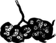
With respect to the mode of action of the secreting cells or nucleated corpuscles, it may be remarked that, in the instance of one of the salivary glands (the sub-maxillary) in the rabbit and the ox, branches of nerve have been traced under the microscope to the individual corpuscles, and it has been found that stimulation of the chorda tympani nerve supplying the gland excites secretion, and that the corpuscles of a gland so excited have a different appearance from those of a gland which has been at rest, losing the sharply defined transparent and slightly striated appearance which they have when at rest, the contour lines becoming indistinct, and the substance altered, so as to be capable of being stained more uniformly with carmine (Heidenhain and Pfluger). So also the secreting cells of the glands of the stomach have been found to have a different appearance during digestion from what they have in an unfed animal, but no appearance of multiplication of the cells at these times can be detected. It therefore appears that the secreting cells act by undergoing, under nervous stimulation, a change of condition, during which they have powers of attraction, elaboration, and transmission of substances. The active condition of the cells is not, however, proved to be in all instances occasional, and excited only by nervous stimulus. While saliva and gastric juice are poured out in response to occasional nervous irritations, the secretions of the kidneys and skin never wholly cease in health; and it is possible that in these organs the corpuscles have a certain amount of continuous activity, comparable with tonicity in muscles.

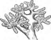
Fig. 34. Lobule of Liver of Oyster.

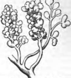
Fig. 35. Lobule of Parotid Gland of Embryo Lamb five inches long.

Fig. 36. Secreting Corpuscles of sub-maxillary gland of the ox with nerve-fibres ending in them. After Pfluger.
Continue to:
