Organs For Receiving Knowledge. Part 2
Description
This section is from the book "The Human Body And Health", by Alvin Davison. Also available from Amazon: The Human Body and Health.
Organs For Receiving Knowledge. Part 2
Deafness
The ear is a very delicate organ, and injury to any of its parts may cause deafness. A sudden pull or a sharp slap on the ear may break the ear drum. In many cases nature will mend this break. Water should be kept out of the ears when diving, by inserting a wad of raw cotton or wool. Closure of the tube extending from the throat to the ear, by catarrh, sometimes causes partial deafness. A growth of germs in the tympanum producing an inflammation as in running ears, may make the three little bones grow together so that they cannot move freely to transmit sound.
More than half of the school children in the United States suffer from some ailment of the ears, eyes, nose or throat. This fact shows the great need of giving proper care to these delicate sense organs. After some diseases, such as scarlet fever, special attention must be given to the ears to avoid partial deafness.
Testing The Hearing
Many children are partly deaf without knowing it. The hearing of every child should be tested occasionally, so that any deafness coming on may be discovered early, when it can be remedied. Each ear should be tested separately by holding a thickly folded handkerchief over the other one. For testing use the tick of a watch or a clock. Children who appear dull often have the best minds, but do not hear distinctly.
Curing Deafness
The deaf and dumb are unable to hear because the ending of the sense organ in the inner ear is imperfect. Some cases of deafness resulting from disease or accident can be cured, but one should never pay attention to the numerous advertisements in newspapers and magazines offering to cure all sorts of deafness. They are the words of quack doctors who rob people of thousands of dollars and are likely to damage the ear seriously. An honest physician does not advertise a sure cure for deafness, but does all in his power to help his patients.
The Sense Of Sight
More knowledge is received through the sense of sight than by all other senses combined. It is a wise provision that we should have two ears and two eyes, for if one is injured the other can do duty for both.
The end organs for sight are the two eyeballs connected behind with the pair of optic nerves leading to the brain.
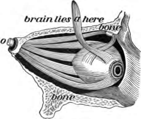
Fig. 127. The eye with its muscles. The side of the skull is cut away, o, the nerve of sight.
The Eyeball
The eyeballs are fixed in soft cushions of fat in the two cavities in the front of the skull. Each eyeball is a globe held in place by six muscles and the optic nerve. The muscles cause the movements of the eyeball.
How The Eyeball Is Protected
The eyebrows on the lower part of the forehead prevent the sweat from running on to the eyeball, and the eyelids keep out dirt and too strong light, and other agents that might cause injury. The eyelashes on the edge of the lids are very sensitive so that the least touch on them causes the eye to be closed.
The tear gland at the upper and outer corner of the eye makes a slightly salty fluid to keep the surface of the eyeball moist and to wash away dust. A tube, the tear duct, carries the tears from the inner corner of the eye into the nose.
Parts Of The Eyeball
The outer covering of the eyeball is a tough coat which is as transparent as glass in front. The transparent part is called the cornea. Just back of the cornea is a curtain, the iris, which has in it a round hole, the pupil, to admit light. The color of the iris gives the brown, black or blue color to the eye.
The outer coat of the eye except the cornea is lined with a thin black membrane, to keep the light from scattering about after it strikes the inner surface of the eyeball. Lying close against this black membrane is a tender pink membrane, known as the retina. This is the real organ upon which the rays of light make an impression to be carried to the brain.
Contents Of The Eyeball
The cavity of the eyeball is filled with three different substances called humors. They are as clear as glass. The middle one and the most firm of all is the crystalline lens, about twice as large and about the shape of a red cinnamon drop. It is held in place just back of the iris by means of a ligament fixed to the black membrane. The cavity in front of this lens is filled with a watery humor while the much larger cavity back of it is occupied by a clear jellylike mass. All these parts of the eyeball may be seen by getting from the butcher or slaughter house the eye of an ox or pig and cutting it open.
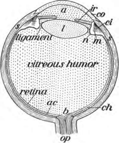
Fig. 128. Lower half of the left eyeball, a, watery humor; b, entrance of the nerve of sight,op; ci, muscles to change the lens for near sight; ir, iris; co, cornea; l, lens; ch, black membrane.
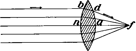
Fig. 129. The rays of light passing through a convex lens bd are brought to a focus at f.
How We See
In order to see an object clearly, the light from it must be brought to a focus on the retina.
The focus is the point at which all rays of light meet as when one holds a burning glass over the hand and brings the rays of the sun together in one tiny spot on the hand. Any transparent substance with outward curving or convex surfaces will bring the rays of light to a focus. The crystalline lens, therefore, brings the rays of light to a focus on the retina. This gives an impression which the optic nerve can carry to the brain.
Continue to:
- prev: Chapter XVIII. Organs For Receiving Knowledge
- Table of Contents
- next: Organs For Receiving Knowledge. Part 3