A Dissection Of The Anterior Surface Of The Adductor Pollicis Manus Muscle
Description
This section is from the book "A Manual Of Dissections Of The Human Body", by R. E. Carrington. Also available from Amazon: A manual of dissections of the human body.
A Dissection Of The Anterior Surface Of The Adductor Pollicis Manus Muscle
Position
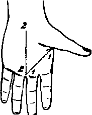
The hand lying upon its dorsal surface, the thumb strongly abducted and rotated outwards.
I. Skin Incisions
1. From the inner border of the base of the proximal Phalanx of the thumb to the inner border of the root of the middle finger.
No. 13.
2. Along the median line from the inner end of the preceding to a point half an inch in front of the wrist.
Reflect the flap backwards and expose the superficial fascia containing granular fat, and
1. Cutaneous branches of artery from the Superficial Palmar arch.
2. Branches of the Musculo-cutaneous nerve over the ball of the thumb.
3. The Cutaneous Palmar branch of the Median nerve at the centre of the wrist.
II
Remove and expose the central thick, and the outer thin portions of the Palmar fascia, and the digitations of the former to the second and third fingers.
III
Remove the preceding and expose
1. The inner tendon of insertion of the Flexor brevis pollicis muscle, and conjoined with it on its inner side the insertion of the Adductor pollicis muscle.
2. The tendon of the Flexor longus pollicis muscle will be seen on the outer side of the preceding, owing to the retraction of the skin.
3. A small piece of the Abductor indicis muscle in front of the Adductor pollicis muscle on the outer side of the index finger.
4. The tendons of the superficial and beneath them of the deep Flexor muscles of the fingers to the second and third digits. They are seen passing into their special sheaths, which extend half an inch into the palm. The Lumbricales muscles of the index and middle fingers are seen, and on the removal of the superficial Flexor tendons are found to arise each from two tendons of the deep.
5. The Princeps Pollicis artery is seen on the thumb, and also its bifurcation.
6. The Radialis Indicis artery on the outer side of the index finger.
7. The outer portion of the Superficial Palmar arch, formed by the Superficial part of the Ulnar artery, joining with the Superficial Volæ branch of the Radial, and a very constant communication with the Radialis Indicis artery. A branch of the arch is seen bifurcating for the contiguous sides of the index and middle fingers. 8. Single branches of the Median nerve to the inner side of the thumb and the outer side of the index finger; and one which bifurcates for the contiguous sides of the index and middle digits. The two inner of these branches are found to supply the two outer Lumbrical muscles. The arteries in the palm are superficial to the nerves.
IV
Remove a. The Superficial Palmar arch and its branches.
b. The branches of the Median nerve to the index and middle fingers.
c. Divide the Flexor tendons of the fingers and pull them forwards. There will now be exposed
1. The Adductor pollicis. muscle arising from the anterior two thirds of the Metacarpal bone of the middle finger, and inserted into the inner side of the base of the proximal Phalanx of the thumb.
2. In front of this, the Abductor indicis muscle on the outer side of the index finger, and the lower part of the Palmar and Dorsal Interossei muscles of the second space; arching over their lower parts the piece of the Transverse Metacarpal ligament between the heads of the second and third Metacarpal bones.
3. The inner fleshy head of the Flexor brevis pollicis, and its tendinous insertion (often containing a sesamoid bone), conjoined with the insertion of the Adductor pollicis muscle.
4. A very small piece of the outer part of the Deep Palmar arch between the Flexor brevis and Adductor pollicis muscles at their inner parts.
5. The Deep branch of the Ulnar nerve supplying the inner head of the Flexor brevis and Adductor pollicis muscles.
Continue to:
- prev: A Dissection To Expose The Flexor Longus Pollicis Manus Muscles
- Table of Contents
- next: A Dissection To Expose The Deep Palmar Arch And Its Branches