Seborrheic Warts
Description
This section is from the book "Skin Cancer", by Henry H. Hazen, A.B., M.D.. Also available from Amazon: Skin Cancer.
Seborrheic Warts
Clinical Course
These lesions, also called senile warts and seborrheic nevi, are very common on the backs and faces of elderly persons. Upon the face they usually develop upon the temples, or sides of the face, or near the midline, especially upon thfe nose. They develop slowly, first appearing as minute yellowish, somewhat scaly, patches, usually roughly oval in shape, and varying in length from 2 to 20 mm. A little later the follicular openings can be recognized as black spots, and still later there is the development of a thick, closely adherent black scale. At first the lesions are but little elevated above the skin level, but later become distinctly so. Itching is rare. The later course has been recently carefully studied by Sutton.7 He concludes that there are three types of advanced lesions- namely, the keratotic, the nevoid, and the verrucose. The keratoid is usually small and is covered with a hard, dark crust. The nevoid approaches the type described by Unna, and appears as a soft, yellowish lesion, with but little horny thickening, while the verrucose lesions have a thin, yellowish crust, which when removed shows a distinctly warty or verrucose condition beneath. None of these lesions disappear spontaneously.
*Engman and Buhman: Medical Herald, 1913, 277.
*Bevan: Quoted by Bloodgood, Progressive Medicine, Dec, 1904, 175. * Bloodgood: Progressive Medicine, Dec, 1904, 175.
*Sutton: Jour. Amer. Med. Assn., 1915.
Pathology
Histologically, these lesions have been studied by Pollitzer,* Unna,* Hartzell,* Fordyce,* Sutton and others. The following account is taken from Sutton's description. The keratoid variety is characterized by great corneus hypertrophy, some parakeratosis, a moderate degree of acanthosis, slight proliferative changes in the germinal layer, with some flattening of the papillary bodies.
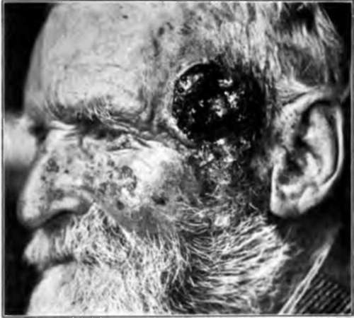
Fig. 3.-This illustration shows a fungous cancer, which sprang from a seborrheic keratosis, similar to the ones upon the cheek and nose. (Sutton's collection).
The sweat ducts are dilated and cysts may be formed. The sebaceous glands are normal, but contain much free fat. The blood vessels of the corium are dilated, and there is some infiltration of the subpapillary portion, with small round and fixed tissue cells.
The nevoid type showed changes similar to those described by Unna in his "nevus seborrheoicus"-that is, but little change in the epithelium, although there is some slight parakeratosis, but a marked increase of pigmentation in the derma, together with dilatation of the blood vessels, and a heavy infiltrate, with round or polygonal cells that resemble nevoid cells. The glandular elements are but little altered.
*Pollitzer: Monatshefte f. prakt. Dermat., 1890, xl, 145. Unna: Histopathology of the Skin (Walkers translation). "Hartzell: Jour. Cutan. Dis., 1903. "Fordyce: Jour. Amer. Med. Assn., 1910, lv, 1624.
The verrucose form is distinguished by considerable hyperkeratosis, very pronounced acanthosis, active proliferative changes in the germinal layer and also at times in other portions of the rete, and most especially by great papillary hypertrophy. The cutis shows changes of a subacute inflammatory nature.
Malignant Degeneration
Basal-celled^eancers are very prone to develop upon these lesions when they are situated upon the face, and occasionally a prickle-celled cancer develops in this situation. When these lesions develop upon the hands, prickle-celled cancer is a common result. The first malignant change noted is a slight ulceration, accompanied by a serous discharge. This ulceration slowly spreads and there is an increase in the induration. It is highly probable that nearly five percent of all these keratoses, especially those of the keratoid variety, terminate in carcinoma.
Treatment
In the early stage these growths are easy to remove. The crust can be removed by an ointment of salicylic acid, and carbon dioxide snow applied to the base for thirty seconds with firm pressure. Another way is to curette off the scale and then use silver nitrate. When a heavy crust has once formed, treatment must be more radical, and the growth should either be excised or treated with the actual or electric cautery. If there is any suspicion of malignancy, operation must be extensive.
Continue to:
- prev: Pigmented Moles
- Table of Contents
- next: Simple Keratosis
Tags
bookdome.com, books, online, free, old, antique, new, read, browse, download