Microscopic Pathology
Description
This section is from the book "Skin Cancer", by Henry H. Hazen, A.B., M.D.. Also available from Amazon: Skin Cancer.
Microscopic Pathology
The tissue can be preserved in any of the accepted fixing agents, although 50 percent alcohol or 10 percent formalin will usually suffice, and is then fixed, blocked, cut, stained, and mounted in the usual manner. Probably, for routine examination, no stain is better than hematoxylin and eosin. A competent microscopist can usually then diagnose the condition without trouble, although one should be selected who knows the difference between the basal- and prickle-celled varieties. In certain growths originating from the glandular appendages of the skin it is sometimes very difficult to decide whether the growths are malignant, and here it is occasionally necessary to have the services of an expert, and then opinions may differ. Usually, however, the question can be decided by any one who has had a broad pathological training. It must always be borne in mind by the novice that inflammatory conditions can cause a great overgrowth of epithelial tissue (Fig. 59), but this should not be mistaken for malignant hypertrophy, for the papillary structure is better retained; the cells of the rete are normal in arrangement and in their relationship to one another, even though they be greatly increased in number. Nor is the basement membrane broken through.
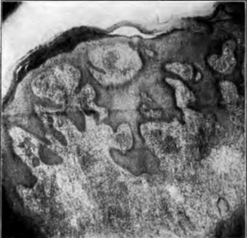
Fig. 69.-In certain chronic infections of the skin, notably tuberculosis, of which this is an example, there is a marked hypertrophy of the epidermis, which may render it difficult to distinguish from cancer. Usually, however, the relationship between the basal- and spino-cells Is not altered. (Author's collection).
The microscopic differences between the squamous- and basal-celled types of cancer has already been fully discussed in the chapters dealing with those subjects.
In general, it may be said that four classes of lesions must be differentiated from the various types of cutaneous cancer-first, those lesions causing nodides, as the benign tumors, cysts of various kinds, chancres, and gummata; second, the ulcerative variety of lesions, as lupus vulgaris, syphilis in the primary or tertiary stage, traumatic ulcers, and certain of the rare granulomata, as sporotrichosis: third, the papillary lesions, such as a more or less diffuse papillomatosis secondary to irritating discharges, exuberant granulation tissue, granulomata pyogenicum, tuberculosis verrucosis, blastomycosis, and very rarely framboesiform syphilis; fourth, lesions causing scarring, as lupus vulgaris, syphilis, lupus erythematosis. and morphea.

Fig. 60.-Chancre of the chin. (Gilchrist's collection).
Continue to:
Tags
bookdome.com, books, online, free, old, antique, new, read, browse, download