Chapter V. Cubo-Celled Carcinomata
Description
This section is from the book "Skin Cancer", by Henry H. Hazen, A.B., M.D.. Also available from Amazon: Skin Cancer.
Chapter V. Cubo-Celled Carcinomata
Origin
It will be remembered that just above the cells forming the basal layer of the rete there is a layer of cuboidal cells, cells somewhat larger than the basal cells, and of a square rather than a columnar shape, and yet with no prickles and not staining as intensely with the acid dyes as do the prickle cells.
Epithelial tumors may arise from these cells, and when they do they possess characteristics of both the basal-celled and squamous-celled carcinomata, or, to be more exact, they form a connecting link between these two radically different types. They have many of the clinical characteristics of the former in that they grow rather slowly, and frequently run a course identical with that of the rodent ulcer, and yet they resemble the more malignant type in that they have a marked tendency to metastasize. At times it is practically impossible to distinguish them histologically from the basal-celled neoplasms, and Bloodgood* thinks that there are undoubtedly many mixed cases.
According to Bloodgood, these tumors were first recognized by Krompecher* in the course of his work on the histology of the basal-celled carcinomata.
Clinical Course
Clinically, these growths arise from pre-existing lesions of the skin, just as do the other varieties of cancer. They may form either fungous or ulcerative lesions, but more usually the former. The tumors often grow very slowly; in a case reported by Bloodgood it took eight years for a diameter of 12 cm. to be reached. In the author's case (Fig. 30) a diameter of 10 cm. was reached in about six months; between these two extremes there are all stages of transition. The fungous neoplasms grow much more rapidly than the ulcers, and metastasis seems to take place sooner. The cancerous tissue resembles anemic granulation tissue, that bleeds easily and is very friable. There is always extreme induration about the edges.
Pathology
The gross pathology is similar to that of the squa-mous-celled cancers, for the alveoli are large, much larger than in basal-celled growths, and contain fine white granular material (cancer cells) that can readily be squeezed out.
*Bloodgood: Progressive Medicine, Dec, 1904. 8 Krompecher: Der Basalzellenkrebs.
When examined under the microscope, it is seen that at first the growths are solid, but that they soon begin to spread out in long finger-like acini, that branch out more or less as they grow downward. The cells resemble large square basal cells, and at times are as large as the prickle cells, but do not contain prickles, nor do they stain with eosin as well as the spinous cells. In addition, there is no tendency for the formation of either whorls or the more advanced epithelial pearls.
Incidence
Out of a total of 997 cases of epithelial tumors, of which 820 were malignant, Bloodgood* reports that there were 5 percent of cuboidal-ceiled tumors. In location, these neoplasms are pretty well scattered. In a study of sixteen cases, Bloodgood states that three were on the lower lip, five on the face, three on the legs, and one each on the scalp, penis, tonsil, tongue, and neck.
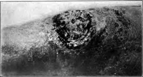
Fig. 30.-This cuboidal-ceiled cancer originated in a varicose ulcer of the leg, and had been present about five months. It has remained well for three years since local removal and cauterization. It would have been safer to have removed the glands. (Author's collection).
Diagnosis
The diagnosis must be made from both prickle- and basal-celled cancers, and in cases of a mixed type of tumor this may be very difficult. The naked eye diagnosis depends on the depth of the malignant infiltration, and the size of the alveoli, and the microscopic diagnosis on the size and character of the cells and the lack of whorls or pearls.
*Bloodgood: Amer. Jour. Med. Sc.. 1914, cxlvii, 76.
Treatment
If caught very early, simple excision with a wide margin will usually suffice to give a permanent cure, but, if the tumor has existed for any length of time, it is absolutely imperative to remove the neighboring lymph glands. Further study of this group of tumors is needed, as we are not yet clear as to when metastasis takes place, although late work by Bloodgood seems to show that this is often comparatively early.
Continue to:
Tags
bookdome.com, books, online, free, old, antique, new, read, browse, download