Pedunculated Adenomata
Description
This section is from the book "Cancer And Other Tumours Of The Stomach", by Samuel Fenwick. Also available from Amazon: Cancer and other tumours of the stomach.
Pedunculated Adenomata
These occur in the form of round, smooth, or lobulated tumours, of a greyish-brown colour and firm consistence, which are attached by short thick stalks to the mucous membrane in the pyloric region. When solitary they may attain the size of an apple, or even of the foetal head at term (Chaput), but if several exist they seldom exceed the dimensions of a walnut and may be as small as peas. In one instance which came under our observation four pedunculated adenomata, each as large as a pigeon's egg, were found attached to the margin of the pyloric ring, and had produced partial obstruction of the orifice. On section the growth is firm and smooth, and sometimes presents several small cysts filled with brownish mucus. Under the microscope it is found to consist of tubular glands, supported by connective tissue and well supplied with blood-vessels, while the mucous membrane which covers it is affected by chronic interstitial inflammation.
It is often impossible to draw a hard and fast line between a large adenoma and an adeno-carcinoma, since the structure may be very similar in the two cases and death may occur in the malignant disease before the formation of metastases. It is also probable that a simple adenoma sometimes assumes a malignant character. Thus, Ferguson has recorded the case of a woman, aged forty-three, who suffered for about a year from pain after food, vomiting, and haematemesis, and presented an ill-defined tumour in the epigastrium. After death the region of the lesser curvature of the stomach was found to be occupied by a large soft growth covered with excrescences, which extended a short distance up the oesophagus and had caused adhesions between the stomach and the under surface of the liver. Although microscopical investigation is said to have proved that the growth was a simple adenoma, the clinical aspect of the case and the naked-eye appearances of the disease clearly indicate that it was malignant in its nature. A similar criticism may be passed upon Lange's case, where a deep ulcer on the anterior wall, surrounded by extensive infiltration, is said to have arisen from the disintegration of a simple adenoma, and as an example of such is usually cited.
(3) Fibromata, like the preceding variety, may be either single or multiple. They usually occur in the pyloric end of the stomach, and are often attached to the edge of the valve. As a rule they are elongated or club-shaped, and measure from one to four inches in length (Bernabei), but they sometimes assume a round or chestnut shape. The surface may be smooth, lobulated, warty, or distinctly villous, and the pedicle is often of the same diameter as the tumour. Microscopically, they either present a papillomatous structure or consist entirely of fibrous tissue covered with thin mucous membrane.
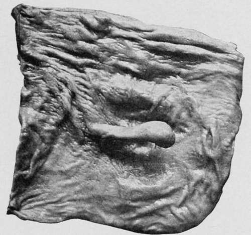
Fig. 62.-Pedunculated fibroma attached to the edge of the pyloric orifice. (London Hospital Museum.).
(4) Pedunculated Lipomata are much rarer than the preceding varieties and are usually solitary. The growth is commonly situated in the central portion of the stomach and on the anterior wall, where it forms a soft lobulated tumour of a pale yellow colour with a short thick pedicle. In one of our cases the tumour measured two inches in length and two and a quarter inches in thickness; while in another it closely resembled a thumb in size and shape. In rare instances a submucous lipoma passes through the muscular coat and forms a pendulous tumour of considerable size beneath the serous investment of the stomach (Orth, Russdorf). Microscopically it is found to consist of fat mixed with fibrous tissue and covered by thin mucous membrane.
(5) Pedunculated Myomata take the form of firm rounded tumours, which vary from the size of a pea to that of a cherry, and are attached to the wall of the stomach by a thin pedicle. They may be single or multiple, and are usually situated in the pyloric region. They consist of unstriped muscle-fibres, which are arranged in a concentric manner and covered by attenuated mucous membrane.
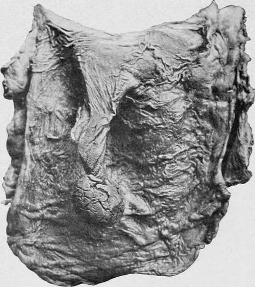
Fig. 63.-A pedunculated fibroma -with a long pedicle (natural size).
Symptoms
The symptoms that accompany polypoid tumours of the stomach vary according to the size and situation of the growth. In nearly one half of the cases where the fundus or central portion of the viscus was affected by mucous polypi, no symptoms whatever were observed during life, while in the rest the patients merely suffered from discomfort after meals, want of appetite, gradual loss of flesh, or from some other indication of disordered digestion. When, however, the pyloric region was involved by the disease, gastric phenomena were almost always present. As a rule the principal cause of complaint was epigastric pain, which was either persistent and unaffected by food, or was only experienced during the period of digestion. Less frequently the patient was subject to sudden and violent attacks, which persisted from a few minutes to several hours and were accompanied by retching and vomiting. In these latter cases there was usually a polypus of considerable length situated near the pylorus, the free extremity of which occasionally prolapsed through the valve and suffered temporary strangulation.
Continue to: