Palpation
Description
This section is from the book "Cancer And Other Tumours Of The Stomach", by Samuel Fenwick. Also available from Amazon: Cancer and other tumours of the stomach.
Palpation
Palpation of the abdomen is a method of examination of the utmost importance. It should be performed, in the first instance, while the patient lies upon his back with his shoulders and head slightly raised and his knees bent over a pillow. The examiner stands or kneels on the right side of the couch, and gently and evenly slides his hand from one part or the abdomen to another, without exercising any sudden or undue pressure with the fingers, which is apt to induce reflex contraction of the recti muscles of the abdomen. For a similar reason care must be taken that the hand is warm before it is applied to the skin. The patient ought not to be permitted to talk, unless it is necessary to distract his attention, and he should be directed to breathe easily and deeply, so as to maintain the stomach in constant movement. Each region is explored in turn, and if any difficulty is encountered in the palpation of the deep structures, the patient should be made to recline upon his left side with the head bent forwards, the knees drawn up, and the back resting against the left knee of the examiner. Sometimes it may be necessary to adopt the knee-elbow position. The points to which particular attention is directed are : (1) The existence of localised tenderness ; (2) thickening of the linea alba and secondary deposits in the skin ; (3) an abdominal tumour.
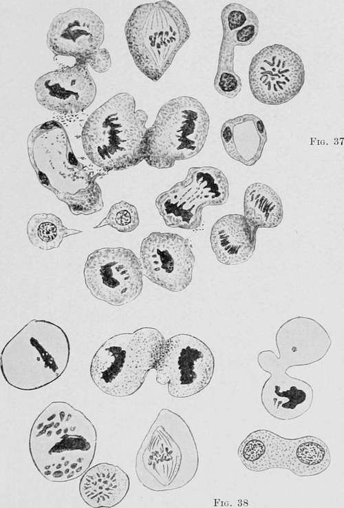
Fig. 37 gives a number of cells in the state of a typical mitoses, taken from parts of the specimen illustrated in fig. 36. To the left side, below the centre, is a mitotic form showing a cell with a huge vacuole and three nuclei; directly opposite, on the right side of the illustration, is a similar smaller cell in which the protoplasm has surrounded a vacuole like a narrow ring. The lowest cell in the figure is an asymmetric mitosis, showing an uneven number of chromosomes and disrupted chromosomes. This is also shown in other cells of fig. 38, particularly in the cells showing a crippled pithode stage. The cells thus pictured were discovered lying detached in the lumen of the gland-ducts as shown in fig. 36. (Hemmeter.).
Continue to:
- prev: Microscopical Examination Of The Contents Of The Stomach. Micro-Organisms
- Table of Contents
- next: Local Tenderness