Sphenoid. Part 3
Description
This section is from the book "The Anatomy Of The Human Skeleton", by J. Ernest Frazer. Also available from Amazon: The anatomy of the human skeleton.
Sphenoid. Part 3
Now examine the position of the spine of the sphenoid. Being on the extreme angle of the wing, it lies immediately in front of the inner end of the Glaserian fissure, and the chorda tympani coming out of the fissure runs on to the surface of the Tensor palati by passing to the inner side of the spine, where it may make a groove (groove of Lucas). Thus the spine has the long ligament fastened to its tip, the chorda tympani on its inner side and the auriculo-temporal nerve passing back between it and the proper capsule of the joint (Fig. 182).
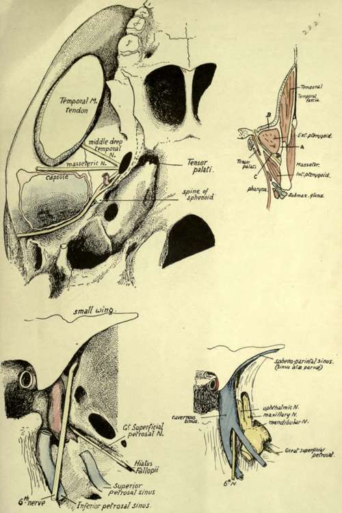
Fig. 182.-The upper left figure shows on the base of the skull the course of some of the branches of the mandibular nerve. A is the basal surface of the great wing, in relation with the upper surface of external Pterygoid, and the middle deep temporal and masseteric nerves are seen crossing this surface, therefore between it and the muscle. The temporal nerve reaches the inferior temporal crest and thus the deep aspect of the Temporal muscle, but the nerve to the Masseter, lying a little further back, passes behind the Temporal tendon (giving a posterior deep temporal nerve to the muscle) and lies along the eminentia articularis, in contact with the articular capsule ; it thus reaches the back part of its muscle. The auriculotemporal nerve runs backwards and outwards, between the sphenoidal spine (long internal ligament) and the capsule (short internal lig.), and then turns out in contact with the back of the capsule, below the Glaserian fissure. The Tensor palati separates the issuing mandibular nerve from the Eustachian tube which issues just internal to the spine. The figure on the right is a scheme of the planes, on coronal section, in the pterygoid region. A is the plane of the deep temporal vessels, B of the (deep) temporal nerve, and C of the inferior dental and lingual nerves. Observe that, as can be appreciated in the previous figure, the external Pterygoid is in relation externally with the Temporal tendon and coronoid process of the mandible: thus the long buccal nerve, piercing the muscle, comes into relation externally with the Temporal and can give an anterior deep temporal nerve to its front part; when it comes forward from under the tendon it appears superficially from under the Masseter, as can be seen from the figure, and lies on Buccinator (see Fig. 206). The lower figures show the relations of nerves, etc., in the region of the foramen lacerum and cavernous sinus. The sinus is removed in one drawing to show the structures deep to it, and its formation is shown in the other. Compare with Fig. 181. Description in text, p. 225.
Again, look at the Eustachian opening : it is situated just internal to the position of the spine, and the tube passes downwards, forwards and inwards from it, and is thus only separated by the Tensor palati from the structures passing through the foramina in the great wing.
The temporal surface articulates by its front edge (Fig. 183) with the malar and is therefore concave from behind forwards as it enters into the formation of the front wall of the temporal fossa as well as its floor. It is covered by and gives origin to the Temporal muscle. Its margin articulates with the malar in front, above this the frontal, the parietal along the top and the squama behind.
The orbital or front surface of the wing, nearly flat, and deepening as it is followed out, forms a great part of the outer or postero-external wall of the orbit, and is in relation with the external Rectus (which gains a small additional origin from its inner part) and the lachrymal vessels and nerve. Its upper and inner border is the lower margin of the sphenoidal fissure and articulates with the frontal outside this; its outer border is practically the front border of the temporal surface (Fig. 183) and carries the malar, while its lower border is separated from the maxilla by an interval, the spheno-maxillary fissure, of which it therefore forms the upper and posterior margin. On the bone this lower border separates the orbital surface from a district of variable size that really belongs to the basal area but is cut off from the pterygoid or true basal region by the pterygoid ridge : this district is spheno-maxillary and will be considered with the pterygoid processes.
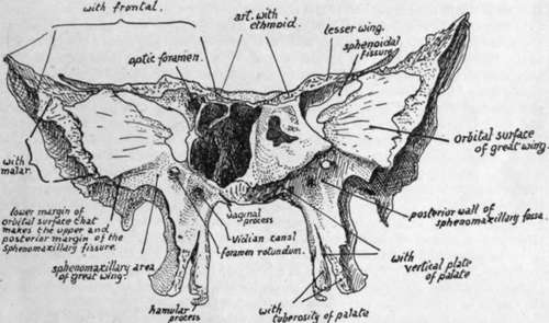
Fig. 183.-Sphenoid and left sphenoidal turbinal from the front.
The small wing or orbito-sphenoid projects outwards and a little forwards from the body. It has a smooth upper surface that is covered by dura mater and forms part of the anterior fossa ; a smooth rounded and concave posterior edge, to which dura mater is attached after it has covered the back of the sphenoidal fissure and which marks the position of the Sylvian fissure, and along which the spheno-parietal venous sinus runs, deep to the dura, to reach the cavernous sinus ; a rough front border that articulates with the frontal; a smooth lower surface which forms the upper boundary of the sphenoidal fissure and, in front of this, the extreme back and inner part of the roof of the orbital cavity.
The posterior margin is continued into the anterior clinoid process, and the widened wing is attached here by two " roots," which enclose the optic foramen between them. The orbital muscles arise from the front and lower surface of the wing round the anterior opening of the optic foramen.
The Pterygoid Processes-each inner process is fused along its front and upper part with the corresponding outer process, but they are separate below. They diverge from each other behind, the plane of the inner plate being practically parallel with that of its fellow, whereas that of the outer plate is nearly at a right angle with the plane of the outer plate of the opposite side. The deep interval thus left between the inner and outer plates behind is termed the inter-pterygoid or pterygoid fossa : its floor is completed below by the tuberosity of the palate bone, which articulates with the front of the two plates in their lower halves and thus closes the gap that exists here between them in the separated bone. A small area is cut off from the upper or proximal part of the pterygoid fossa by an oblique ridge, and constitutes the scaphoid fossa : the front part of the Tensor palati arises here, and its area of origin extends from this backwards and outwards along the margin of the great wing internal to the foramina, nearly as far as the spine of the sphenoid (Fig. 182). Otherwise the pterygoid fossa is occupied by the Internal Pterygoid muscle, which arises from the inner side of the outer plate and from the tuberosity of the palate between the plates. The External Pterygoid takes origin from the outer side of the outer plate, an area continuous with that on the basal surface of the great wing. Thus the external plate may be looked on as a muscular process, probably developed only in association with such function ; but the inner plate is a bone developed in the pharyngeal wall and only gives origin to one muscle, the upper Constrictor of the pharynx, which arises from the lower half of its posterior border. It is covered by mucous membrane on its inner surface, where it forms the outer wall of the posterior opening of the nasal fossa, and its outer surface is in relation with Internal Pterygoid and Tensor palati, but does not give origin to them. Its lower end is prolonged into a hooked hamular process directed backwards and outwards and showing a deep notch on its outer and front aspect, round wlrch the tendon of Tensor palati turns in to enter the soft palate.
Continue to: