Sphenoid
Description
This section is from the book "The Anatomy Of The Human Skeleton", by J. Ernest Frazer. Also available from Amazon: The anatomy of the human skeleton.
Sphenoid
A single bone situated in the base of the skull, but reaching the side walls : consisting of a centrally placed body from which two wings, great and small, project on each side, and two pterygoid plates project downwards on each side, the inner from the body and the outer from the under surface of the great wing.
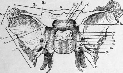
Fig. 179.-Sphenoid seen from above and behind. A., marginal articulation with ethmoid ; B., with frontal ; C, with parietal; D., with squama of temporal, a.. Small wing ; b., great wing ; C, sphenoidal fissure ; d., jugum sphenoidale ; e., optic groove ; (., olivary eminence ; g., optic foramen ; h., anterior clinoid process ; »'., posterior clinoid process; k., dorsum sellae ; foramen rotundum ; m., carotid groove ; n., lingula ; o., foramen ovale ; p., foramen spinosum ; r vidian foramen.
The body, on the whole cuboidal, articulates behind with the occipital, with which it is synostosed about twenty-five, and in front carries the ethmoid : it is hollowed out by the sphenoidal air sinuses * which extend into it from the nasal fossae within a few years after birth, and when fully developed occupy practically the whole of the body and may extend even into the attached parts of the wings and pterygoid processes. The two sinuses are separated by a septum, which may be incomplete and is usually asymmetrical : there are as. a rule two partial septa also present, one in each sinus near its upper and outer part. The sinuses are partly closed by two sphenoidal turbinals* thin paper-like bones, that have openings above their centres through which the cavities are connected with the ethmo-sphenoidal recesses in the nasal fossae.
* Tor further details, see p. 249 (sphenoidal sinus), and p. 227 (sphenoidal turbinals).
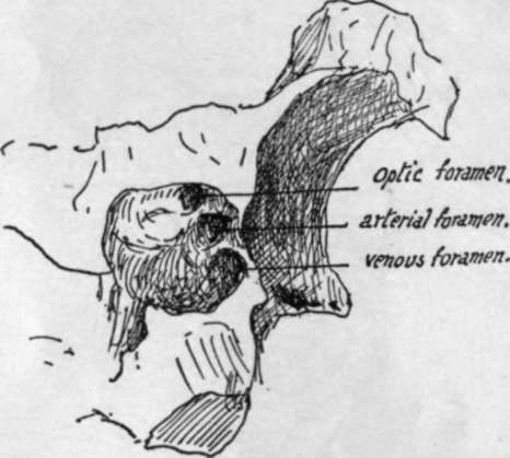
Fig. 180.-A specimen in which the ligamentons bands joining the three clinoid processes have ossified, with the result that foramina exist which are not present with bony boundaries in the sphenoid as usually seen. The carotid passed through the arterial foramen and the circular sinus through the venous foramen. A carotid foramen is not uncommon ; the double structure is more rare.
The upper surface of the body presents a deep hollow open at the side, the pituitary fossa, for lodging the pituitary gland and its surrounding vascular plexus ; in front of this the olivary eminence separates it from the optic groove, which runs into the optic foramen at each side. In front of the groove the flatter jugum sphenoidale stands at a higher level than the rest of the body and is continued laterally into the lesser wings.
The optic chiasma is attached to the front of the infundibulum of the pituitary, and is therefore raised above the level of the bone and does not lie in the optic groove, but the optic nerves run from it into the optic foramina accompanied by the ophthalmic arteries. The pituitary fossa is roofed in by the diaphragma sella, a curtain of dura mater that is pierced centrally by the stalk of the gland. The diaphragm (Fig. 181) is attached to the front and back margins of the fossa, and at the sides to the clinoid processes and interclinoid ligaments : the an erior clinoid process is a thick angular projection from the base of the small wing towards the fossa, and the posterior process is the prominent upper and lateral angle of the dorsum sella that overhangs the back of the fossa. The middle clinoid process, if present, is a low point of bone situated between and below the other two at the side of the fossa. Interclinoid ligaments connect these processes.
At the sides, where it slopes down to be continuous with the great wings, the body supports the cavernous sinus on each side (Fig. 182), and the system of venous spaces (known as the circular sinus) that surrounds the pituitary body joins the two cavernous sinuses. The internal carotid artery reaches the bone here, deep to the s'nus, from the foramen lacerum (see Complete Base), and runs along it in the carotid groove, to turn in under the overhanging anterior clinoid process and then up to the inner side of the process behind the optic nerve ; here it gives its ophthalnvc branch. Thus the artery passes in front of the ligamentous connection between the anterior and middle clinoid processes, which may be ossified, so that there may be foramina at each s:de of the upper surface of the body, as shown in Fig. 180, for the optic nerve, the carotid artery, and the circular sinus.
The lower surface of the body has a thick, wedge-shaped bar running along its front part, supporting the septum between the air sinuses above and ending in front in the prominent rostrum : the hinder end of this bar of bone is continuous with the rest of the body and is partly overlapped by the vaginal processes that turn in from the internal pterygoid plates. This aspect of the body, as can be seen in the skull, carries the vomer, and the two alas of that bone open out below the sphenoidal body and fit in under cover of the vaginal processes. Behind this region the under surface is lined by mucous membrane of the roof of the naso-pharynx.
On each side the body is continuous with the great wing : the carotid groove commences on the back of this junction, and here there is a projection on the outer side of the groove known as the lingula. This appears to be only present as a guard for the artery, but its morphological value is doubtful ; it has a separate centre of ossification, and is laid down as a separate cartilaginous structure in the embryonic skull.
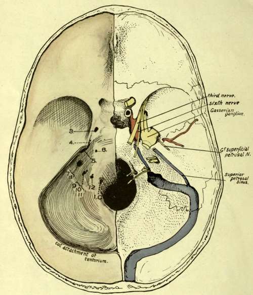
Fig. 181.-Cranial surface of base of skull, with dura mater in position on the left side, the tentorium cere-belli being cut away. 3-12, foramina made by the corresponding cranial nerves piercing dura mater ; 1. C. first cervical nerve foramen. Observe that when the dura is in position the margins of the foramen magnum are not defined, owing to the continuation of the dural slope on to the odontoid, consequently the foramina have the appearance of being higher than they really are in the skull. The membrane covers in the pituitary fossa, forming the diaphraqma sella, attached to the inter-clinoid ligaments. The structures represented on the right side are sufficiently described in the text. Compare with Fig. 182 and p. 225.
Continue to:
- prev: The Petrous
- Table of Contents
- next: Sphenoid. Part 2