Lachrymal
Description
This section is from the book "The Anatomy Of The Human Skeleton", by J. Ernest Frazer. Also available from Amazon: The anatomy of the human skeleton.
Lachrymal
Small shell-like bones placed near the front part of the inner orbital wall. Each bone articulates by its posterior margin with the os planum of the ethmoid, by its front border with the nasal process of the maxilla, rests below on that bone, and reaches the frontal above : its deep surface covers the antero-lateral aspect of the lateral mass of the ethmoid where the os planum is deficient, and forms outer walls here for the anterior ethmoidal cells and the infundibulum and a small part of the wall of the middle meatus, and a prolongation downwards of this surface, making the inner wall of the nasal duct, meets and articulates with an upper process of the inferior turbinate bone which forms the lower end of this inner bony wall. This downward process of the lachrymal also articulates by its posterior part with the uncinate process of the ethmoid, and to a very small extent helps to close in the opening of the antrum of Highmore.
The deep surface shows impressions for the cells that are walled in by it.
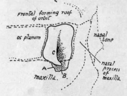
Fig. 189.-Orbital aspect of right lachrymal bone, with the adjoining bones indicated by interrupted lines. A., process downwards forming inner wall of nasal duct and joining inferior turbinate; B., hamular process; C, lachrymal crest.
The superficial, external, or orbital surface, on the whole slightly concave, has a posterior part level with the os planum, separated by a lachrymal crest from the deep fossa that holds the lachrymal sac and is continued down into the nasal duct. The crest ends below in the hamular process, turning forwards and outwards and resting on the maxilla, and making the outer margin of the opening. In rare instances the hamular process is found extending forward to the orbital margin ; on the other hand, the process may be absent. The Tensor tarsi arises from the crest and passes forward to the outer side of the nasal sac, which it is supposed to compress on its contraction.
The bone is sometimes perforated, and is occasionally represented by several ossicles. Its complete absence has been recorded.
Ossification commences in membrane before the end of the third month, on the surface of the nasal capsule. It reaches the frontal within a few weeks, but does not extend back to articulate with the os planum until some months later, as the posterior or ethmoidal portion of the bone is the last to be formed.
Continue to:
- prev: Ethmoid
- Table of Contents
- next: Vomer