Palate Bone
Description
This section is from the book "The Anatomy Of The Human Skeleton", by J. Ernest Frazer. Also available from Amazon: The anatomy of the human skeleton.
Palate Bone
Each palate bone consists of : (1) a vertical plate, which is applied to the posterior part of the inner surface of the body of the maxilla ; (2) a horizontal plate, which projects nwards from the lower end of the vertical plate, lies in the plane of the palatine process of the maxilla, and articulates mesially with its fellow of the opposite side ; (3) a tuberosity that projects downwards, outwards and backwards from the vertical plate, and so lies behind the lower part of the back of the maxilla and separates it from the pterygoid plates of the sphenoid, against which it is placed.
The vertical plate is not only applied to the inner side of the maxilla, but extends back behind the level of this bone to articulate with the front border of the internal pterygoid plate, and in this way its hinder part forms the inner wall of the sphenomaxillary fossa. These two articulations cause a difference in the direction of the back and front portions of the vertical plate : the front part (Fig. 185), being applied to the maxilla, turns outward at its upper end with the maxillary surface, and thus lies between the maxilla and the ethmoid and reaches the orbit, where it forms part of the floor ; whereas the posterior part, following the margin of the internal pterygoid plate, turns in at the top (Fig. 183) and comes to lie below the body of the sphenoid. The vertical plate can be described, therefore, as terminating above in an anterior orbital process, directed upwards and outwards, and a posterior sphenoidal process, directed upwards and inwards. Between these two is a deep sphenopalatine notch, converted into a foramen by the sphenoidal turbinate that lies above it. Sometimes a large orbital process may convert the notch into a complete foramen : this has also been seen double.
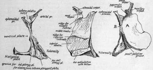
Fig. 187.-Right palate bone. 1. Posterior aspect. Observe the deep groove for internal pterygoid plate against which the bone is placed. 2. From the nasal or inner side. 3. From the outer side. The greater part, A., of this surface rests against the maxilla, but a small part, B., projects back beyond that bone and forms the inner wall of the spheno-maxillary fossa ; the post. pal. canal runs down from this surface, between A. and the tuberosity.
The orbital process, very variable in size, has :-
(a) A maxillary surface, applied to the maxilla ;
(b) an ethmoidal (upper) surface, which supports the ethmoid and shows a crest for the middle turbinal, a part of the ethmoid ;
(c) a spheno-maxillary (or zygomatic) surface, looking outwards and backwards behind the maxilla and therefore into the spheno-maxillary fossa ;,
(d) an orbital (terminal) surface, visible between ethmoid and maxilla in the floor of the orbit.
The sphenoidal process has :-
(a) An outer or spheno-maxillary surface ;
(b) an inner or nasal surface ;
(c) an upper terminal part applied to the sphenoidal body. It is grooved behind by the margin of the internal pterygoid plate.
The vertical plate below the processes and notch has an inner surface that is purely nasal, crossed by a rough ridge that carries the lower turbinate bone, and an outer surface that is mostly maxillary, but has a small triangular area behind this and immediately below the notch (Fig. 187) which forms the inner wall of the spheno-maxillary fossa. Below this the tuberosity begins to grow out from the bone. On this surface of the plate, in front of the tuberosity, the posterior palatine groove is seen running downwards and forwards : this part of the bone is applied to the maxilla, which therefore completes the canal. The lower part of the groove may be partly or completely closed in by the tuberosity.
The horizontal plate has rough articular front and inner margins for the maxillary palate and its fellow respectively, a concave posterior edge to which the palatine aponeurosis (soft palate) is attached, with a prominent posterior nasal spine centrally, where the Azygos uvulae muscle arises, a rough lower surface, and a smooth upper or nasal surface which forms a prominent median crest with its opposite fellow, for the support of the vomer.
The tuberosity extends in a direction downwards and outwards and slightly backwards. It has a rough antero-external maxillary surface, separated by the posterior palatine groove from the maxillary surface of the vertical plate, and a posterior surface which is applied to the front of the pterygoid processes. This surface (Fig. 187) shows a deep groove internally for the internal pterygoid plate, a rough articular area obliquely directed by its outer margin for the outer plate, and a shallow concave fossa between these that completes the floor of the pterygoid fossa (see Fig. 161) and gives origin to fibres of the Internal Pterygoid muscle. This origin extends on to the lower surface (Fig. 161) of the tuberosity and from this runs on to the outer part, which is visible between the external plate and maxilla.
This origin of part of Internal Pterygoid gives its front and lower fibres the appearance of being superficial to the lower fibres of the External Pterygoid.
Articulations
With the maxilla (vertical plate, horizontal plate and tuberosity), ethmoid and inferior turbinal (vertical plate), sphenoid (vertical plate and tuberosity), vomer and its fellow (horizontal plate).
Development
The bone is developed in membrane on the inner aspect of the cartilaginous nasal capsule, from a centre that appears during the eighth week in the region of the future tuberosity ; from this ossification extends into the palate fold as this meets its fellow, to form the horizontal plate, and more slowly upward to make the vertical plate. Owing to the relative small vertical measurement of the nasal cavities at birth, associated with the incomplete growth of the maxillae, the palate bone at this time shows a vertical plate of only about the same length as the horizontal plate.
Continue to: