Operation Of Tying The Radial Artery
Description
This section is from the book "Anatomy Of The Arteries Of The Human Body", by John Hatch Power. Also available from Amazon: Anatomy of the Arteries of the Human Body, with the Descriptive Anatomy of the Heart.
Operation Of Tying The Radial Artery
The radial artery may be tied in the upper part of the fore-arm by making an incision over the interval between the pronator radii teres and supinator longus. In the lower part of the fore-arm it will be found between the flexor carpi radialis and supinator longus. In either case, after making the incision through the integuments, the fascia should be divided on a director: a small vein will be found on either side of the artery, and the radial nerve will lie on its external side. The possibility of mistaking the superficialis volae for the trunk of the radial should be borne in mind. In judging of the strength of the pulse at the wrist, it will be necessary to attend to the deviations in the course and size of the radial artery.
Sir Philip Crampton succeeded in curing a circumscribed traumatic aneurism of the radial artery as it passes behind the wrist, by the application of Dr. Carte's compressing instrument upon the artery leading to the tumor.*
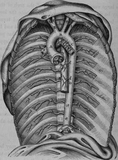
Fig. 33. The Thoracic Aorta and its Branches.
A, Ascending portion of the Arch of the Aorta. B, Middle portion of the Arch. C, Termination of the descending portion of the Arch. D, Thoracic Aorta. E, Arteria Innominata, or Brachiocephalic Artery. F, Right Common Carotid Artery. G, Right Subclavian Artery. H, Left Common Carotid Artery. I, Left Subclavian Artery. K. K, Inferior Phrenic or Diaphragmatic Arteries, which in this case came abnormally from the Coeliac Axis, a, a, a, Sigmoid or Semilunar Valves of the Aorta, b, Origin of the Right Coronary Artery, c. Origin of the Left Coronary Artery, d. Right Bronchial Artery, in this case arising from the concavity of the Arch of the Aorta, e, Left Bronchial Artery, having a similar origin, f, CEsophageal Arteries, g, g. g. g, g, g, Left Inferior or Aortic Intercostal Arteries, h. h, h. h. h, h. Right Inferior or Aortic Intercostal Arteries. 1, Trachea. 2, Right Bronchus. 3, CEsophagus. 4, 4, Portion of the Diaphragm.
Continue to: