The Radial Artery
Description
This section is from the book "Anatomy Of The Arteries Of The Human Body", by John Hatch Power. Also available from Amazon: Anatomy of the Arteries of the Human Body, with the Descriptive Anatomy of the Heart.
The Radial Artery
The Radial Artery, smaller than the ulnar, but more in the direction of the brachial artery, descends towards the wrist, being related posteriorly, from above downwards, to the tendon of the biceps, the insertion of the supinator brevis, the pronator teres, the radial origin of the flexor sublimis, the flexor pollicis longus, and the pronator quadratus muscles: externally, it is related to the supinator longus muscle, which overlaps it a little; and internally to the pronator teres above, and flexor carpi radialis lower down. Anteriorly it is covered only by fascia, integuments, and the approximation of the muscles at either side. Thus in the upper part of its course the artery will be found between the supinator longus and pronator teres, whilst below this it lies between the supinator radii longus and flexor carpi radialis. The radial artery is accompanied by two veins, the venae comites, and in the two superior thirds of the fore-arm by the radial branch of the musculo-spiral nerve, which lies to its outer or radial side : below this point the nerve forsakes the artery and winds round the outside of the radius, passing underneath the tendon of the supinator radii longus, in order to arrive at the outer side of the posterior part of the fore-arm. At the lower extremity of the fore-arm the artery turns round the external lateral ligament of the wrist-joint, being parallel to the radial extensor muscles, and covered by the extensor muscles of the thumb. Here it pierces the abductor indicis manus muscle, and terminates, in crossing the palm of the hand, under the name of the palmaris profunda. As the artery is passing obliquely across the back of the outer portion of the wrist, it will be found lodged in a triangular space, the base of which corresponds to the back part of the lower extremity of the radius; the apex is situated at the metacarpal bone of the thumb; one side is formed by the extensor secundi internodii pollicis and extensor carpi radialis longus; and the other, or radial side, is formed by the tendon of the extensor primi internodii pollicis. Immediately underneath the integuments covering this hollow space, we find the origin of the radial vein and some branches of the radial division of the musculo-spiral nerve.
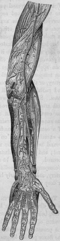
Fig. 29. Represents the Deep Arteries of the upper Extremity.
A, A, Brachial Artery. B, B, Radial Artery. C, C, Ulnar Artery. D, D, Auterior Interosseal Artery. K, Slender twig to the Brachialis Anticus. P, Deep Palmar arch of Arteries formed by the Communicans Profunda of the Ulnar, and Palmaris Profunda of the Radial Arteries. Q, Portion of First Dorsal Interosseous Muscle. 1, Coraco-brachialis Muscle. 2, Long portion of Triceps Muscle. 3, Brachialis Anticus. 4, Internal Intermuscular Septum. 5, Short portion of Triceps Muscle. 6, Extensor Carpi Radialis Longus. 7, Twig to the Brachialis Anticus. 8, Part of the origin of Pronator Radii Teres. 9, Origins of Flexor Carpi Radialis and Palmaris Longus. 10, Extensor Carpi Radialis Brevis Muscle. 11, Supinator Radii Brevis Muscle. 12. Portion of the Flexor Profundus Muscle. 13, Insertion of Pronator Teres cut. 14, 15, Flexor Pollicis Longus having the Radial Artery passing over it. 16, 16, 16. The Interosseous Ligament with Anterior Interosseal Artery. 17. Pronator Quadratus with branch of Interosseal Artery. 18, Anastomosis between Anterior Interosseal, the Deep Palmar Arch, and the Anterior Carpal Arteries. 19, 20, Abductor Minimi Digiti Muscle. 21. 21, 21, Palmar Interossei Muscles, a, Muscular Branch, c, Superior Profunda Artery, d, Inferior Profunda Artery, e, f. g, h, Muscular branches to Triceps and Brachialis Anticus Muscles, i. Anastomotic Artery. 1, Radial Recurrent Artery, m, Superficialis Volae cut. n, Princeps Pollicis Artery, o. Anterior Ulnar Recurrent Artery ascending to anastomose with the Anastomotic artery, r, r, r, Digital Arteries, s, s, s. Cut ends of the Digital Arteries of the Superficial Palmar Arch, t, t, t, t, u, u, u, u, v, v, v, v, Anastomoses between the Digital Arteries.
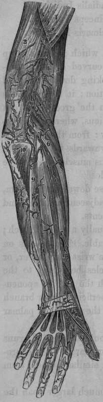
Fig. 30. Represents the arteries of the posterior part of the upper extremity which are seen after the removal of the skin and aponeurosis.
1, Deltoid Muscle. 2, Triceps Extensor Cubiti. 3, Biceps Flexor Cubiti. 4, Brachialis Anticus. 5, Supinator Longus. 6, Extensor Carpi Radialis Longus. 7, Extensor Carpi Radialis Brevis. 8, Extensor Communis Digitorum. 9, Extensor Carpi Ulnaris. 10, Anconeus Muscle. 11, Flexor Carpi Ulnaris. 12, Extensor Osss Metacarpi Pollicis. 13, Extensor Primi Internodii Pollicis, a, a. a, Muscular branches of the Superior Profunda, b, Branch of the Superior Profunda, c, c, Anastomoses between the Superior Profunda and Twigs from the Interosseal and Posterior Ulnar Recurrent Arteries, d, Twig from the Radial Recurrent Artery, e. Twigs from the Interosseal Artery, f, Twig from the Interosseal Artery, g, h. Arterial Anastomosis, i, Radial Artery, k. k, k, k, Twigs from the Anterior Digital Arteries to the backs of the lingers.
The branches of the radial artery are the following:
Radial Recurrent.
Muscular.
Metacarpal.
Superficialis Volae.
Anterior Carpal.
Princeps Pollicis.
Posterior Carpal.
Palmaris Profunda.
The Radial Recurrent
This branch, which arises high up in the fore-arm, proceeds at first in a curved direction outwards, the convexity of the curve looking downwards and lying below the radio-humeral articulation: it then ascends on the front of the supinator brevis, in the groove between the supinator longus and brachialis anticus, where it anastomoses with the superior profunda artery: from the convexity of its arch it sends many branches downwards to be lost in the supinator brevis and supinator longus muscles, and in the upper extremities of the extensor muscles.
The Muscular Branches
In its course down the fore-arm, the radial artery sends branches to the adjacent muscles, and through the aponeurosis to the integuments.
The Superficialis Volse
This is usually a small branch; sometimes, however, it is very considerable. It descends on the front of the annular ligament of the wrist; then over, or through the origins of the small muscles belonging to the thumb. It next turns inwards, beneath the palmar aponeurosis, and, by anastomosing with the superficial palmar branch of the ulnar artery, contributes to form the superficial palmar arch already described.
The Anterior Carpal Artery
The Anterior Carpal Artery is small but constant. It runs transversely inwards, along the inferior margin of the pronator quadratus, to anastomose with a similar branch from the ulnar.
Continue to: