The Brachial Artery
Description
This section is from the book "Anatomy Of The Arteries Of The Human Body", by John Hatch Power. Also available from Amazon: Anatomy of the Arteries of the Human Body, with the Descriptive Anatomy of the Heart.
The Brachial Artery
This artery is a continuation of the axillary: it commences opposite the lower margin of the teres major and latissimus dorsi tendons, passes obliquely downwards and outwards, and terminates nearly opposite the coronoid process of the ulna: on the removal of the integuments, the artery will be found lying under cover of the brachial aponeurosis. After the aponeurosis has been removed, the vessel will be seen overlapped by the fleshy belly of the coraco-brachialis muscle, then by the biceps muscle, and still lower down covered by the semilunar fascia derived from the tendon of the biceps : these are its anterior relations : internally it is related, in addition to the integuments and fascia, to the basilic vein, to the inferior profunda artery, and to the ulnar and internal cutaneous nerves; externally it is related to the coraco-brachialis and biceps muscles, and to an areolar interval placed between the biceps and brachialis anticus: posteriorly it corresponds first, to the triceps muscle, from which it is separated by the superior profunda artery and musculo-spiral nerve; next it rests on the insertion of the coraco-brachialis muscle; and, in the remainder of its course, it lies upon the brachialis anticus.
The brachial nerves surround the artery, and are related to it in the following order: behind it, but accompanying it merely for a short distance, is the musculo-spiral nerve : the external cutaneous nerve at first descends along its outer side, separating it from the coraco-brachialis muscle; but lower down, it inclines outwards, perforates the last-named muscle, and loses its relation to the artery. The internal cutaneous nerve lies at first on the inside of the artery, being situated on the front of the ulnar nerve, which it consequently separates from the median: lower down, the branches of the internal cutaneous nerve become superficial, and one principal filament covers the artery at its termination. The ulnar nerve descends on the inside of the vessel, but towards the middle of the humerus separates from it, and inclines still more internally, and accompanies the inferior profunda artery; and lastly, the median nerve lies on the outside of the brachial artery above; but lower down, at about the junction of the lower with the two upper thirds of the arm, it crosses the artery, usually over its anterior surface, in order to arrive at the inner side of the vessel. The veins accompanying the artery are two in number, and are termed venae comites: about the middle of the arm they unite with the basilic vein, which usually perforates the brachial aponeurosis in this situation. Such are the relations of the brachial artery in its course down the arm. At its termination it sinks into a triangular space, in front of the elbow-joint, bounded on the outside by the supinator radii longus, and on the inside by the pronator radii teres muscle; the latter muscle overlapping the artery in this situation. In this space it lies on the brachialis anticus muscle, having the tendon of the biceps to its outside, the median nerve to its inside, while in front it is covered by an aponeurotic slip of a semilunar form, sent downwards and inwards from the tendon of the biceps muscle to join the anti-brachial aponeurosis a little below the internal condyle: this is called the semilunar fascia of the biceps; its upper margin is concave and directed upwards and inwards; its insertion into the fascia of the fore-arm is much broader than its origin from the tendon of the biceps.
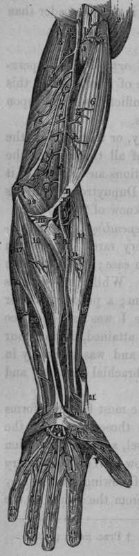
Fig. 28. Represents the Arteries of the upper ex-tremity, which are seen when the skin and fascia have been removed.
A A, Brachial or Humeral Artery. B B, Radial Artery. C, Ulnar Artery. K, Muscular branch to the Brachialis Anticus.
0, Superficialis Volae Artery. P P, The Superficial Palmar Arterial Arch, formed by the Ulnar and Superficialis Volae Arteries. Q, Digital Artery of thumb. S, Twig to the Palmaris Brevis Muscle. V, Princeps Pollicis Artery, running along the internal margin of the thumb, a, Twig to the Triceps, b, Small branch to Coraco-brachialis and Biceps, c, Superior Profunda about to enter between the two portions of the Triceps, d. Inferior Profunda, arising opposite the insertion of the Coraco-brachialis Muscle, e, f, Muscular Branches, g, h, Small twigs to the Biceps, i, The Anastomotic Artery. 1, Radial Recurrent Artery, m, Twig to the Pronator Teres and Flexor Carpi Radialis Muscles, n, Branch to the Supinator Radii Longus. r, The Radialis Indicis Artery, t, t, t, t, The four Digital Arteries, u, u, u, u, The arches formed by the Digital Arteries.
1, Portion of Pectoralis Major. 2, The Deltoid Muscle. 3, Upper portion of Biceps Muscle. 4, Coraco-brachialis. 5, Triceps. 6, Belly of Biceps. 7, Internal Intermuscular Septum. 8, Short portion of Triceps. 9, Brachialis Anticus. 10, Tendon of Biceps. 11, Semilunar Fascia from Biceps Tendon. 12, Pronator Teres. 13, Internal Condyle. 14, Supinator Radii Longus Muscle. 15, Pronator Teres crossed by Radial Artery. 16, Flexor Carpi Radialis. 17. Palmaris Longus. 18, Flexor Carpi Ulnaris. 19, Extensor Carpi Radialis Longior. 20, Portion of Flexor Digitorum Sublimis, or Perforatus. 21, Extensor Primi Internodii Pollicis. 22, Extensor Ossis Metacarpi Pollicis. 23, Palmar Aponeurosis. 24, Tendons of the Superficial Flexor, crossed by the Superficial Palmar Arch of Arteries.
The Operation Of Tying The Brachial Artery
This operations may become necessary for the cure of aneurisms of this vessel, or in consequence of a wound inflicted on it or upon the radial, ulnar, or interosseous arteries.
Continue to: