Veins Of The Arm And Fore Arm
Description
This section is from the book "Anatomy Of The Arteries Of The Human Body", by John Hatch Power. Also available from Amazon: Anatomy of the Arteries of the Human Body, with the Descriptive Anatomy of the Heart.
Veins Of The Arm And Fore Arm
Before proceeding with the dissection of the brachial artery the student is recommended carefully to examine the superficial veins of the arm and fore-arm; for this purpose he should remove the integuments from off the front of these parts, when the veins and superficial nerves will be exposed lying between the skin and fascia.
Venisection is usually performed at the bend of the elbow, because there are in this situation a number of superficial veins, easily made prominent and easily compressed. On the outside of the bend of the elbow we observe the cephalic vein, ascending, having derived its principal origin from the cephalic vein of the thumb. On the inside we see the basilic vein, which seems to be a continuation of the small vein of the little finger, termed vena salvatella. On the middle line of the front of the fore-arm we see the median vein, which, as it approaches the elbow-joint, divides into an internal and external branch : the internal branch is the median basilic vein ; it crosses in front of the brachial artery at a very acute angle, being separated from it immediately beneath the bend of the elbow, by the semilunar process of the biceps tendon, called also the semilunar fascia of the biceps : some of the branches of the internal cutaneous nerve pass in front of it, and others behind it. The external branch, smaller than the internal, is termed the median-cephalic vein ; it ascends obliquely upwards and outwards, in front of the trunk of the external cutaneous nerve, to join the cephalic vein. The basilic and cephalic veins, being thus reinforced, ascend in the arm, the former along the internal and the latter along the external margin of the biceps muscle. The basilic vein unites with the venae comites of the brachial artery, and the large vessel formed by their union becomes the axillary vein.
In the middle of the fore-arm, near the bend of the elbow, the median vein, before it gives off its median basilic and median cephalic veins, receives at its posterior surface, from the deep-seated parts of the fore-arm, a vein called the me-diana profunda.
When the operations of venaesection is determined on, the student will observe that the median basilic is the vein which presents itself most prominently; and, if this be selected for the operation, great caution will be necessary, in order to avoid wounding the brachial artery, which lies beneath it. On this account the student is advised to select the median cephalic vein in preference, at all events until he has become somewhat expert in performing the operation. A wound of the artery during venaesection may be denoted by the blood issuing in jerks, and being of a bright arterial color. These appearances may exist, however, without any such wound, and therefore need not always excite alarm: on the contrary, the artery may be punctured without any particular symptom to indicate the accident. When there is reason, from the great force with which the blood is projected, to suspect that this accident has occurred, and there is no pain, swelling, nor effusion present, we may apply a graduated compress, keep the limb quiet, and wait the result, which may be various. Sometimes the wounded vessel may heal without any unpleasant consequence: in other cases, the external wound of the vein is healed, but the wound in the posterior wall of the vein may form an adhesion with the wound in the anterior wall of the artery, and thus there remains a direct communication between the artery and vein. When this direct communication exists between the two vessels, the affection is termed aneurismal varix; but if the areolar tissue intervening between the two vessels has been distended into the form of a sac, which establishes a medium of communication between the artery and vein, then the disease is termed varicose aneurism. The latter is the more serious, as it may terminate in aneurism of the artery; but it is seldom that either of them requires any operation.
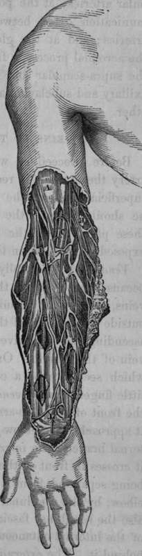
Fig. 27. Represents portion of the Surgical Anatomy of the Fore-arm.
A, Fascia over the Biceps Muscle. B, Basilic Vein and Internal Cutaneous Nerve. C. Brachial Artery and the Venae Comites. D, Cephalic Vein and External Cutaneous Nerve coming out from behind it. E, Median Cephalic Vein and a communicating vein to the Venae Comites. F. Median Basilic Vein. G, Radial Artery. H. Lymphatic Gland. I, Radial Artery seen through "an opening made in the fascia. K. Ulnar Artery and Ulnar Nerve. L, Palmaris Brevis Muscle.
The student may now remove the veins and brachial aponeurosis, so as to expose the brachial artery.
Continue to: