The Thoracic Aorta
Description
This section is from the book "Anatomy Of The Arteries Of The Human Body", by John Hatch Power. Also available from Amazon: Anatomy of the Arteries of the Human Body, with the Descriptive Anatomy of the Heart.
The Thoracic Aorta
This great division of the descending aorta may be said to commence opposite the third dorsal vertebra, and to terminate in passing between the pillars of the diaphragm. As far as the tenth dorsal vertebra it is situated in a region called the posterior mediastinum: this region approaches somewhat to the form of a prism, and extends from about the third to the tenth dorsal vertebra: its sides are formed by the two pleurae; its apex is situated anteriorly and corresponds to the back part of the pericardium, and its base is formed by the bodies of the vertebrae from the third to the tenth. The direction of the thoracic aorta is downwards, forwards, and to the right side. Its posterior surface rests on the spine and demi-azygos vein, and usually on the third, fourth, and fifth intercostal veins of the left side : the intercostal arteries arise from this part of the vessel. Its anterior surface is covered by the root of the left lung, by the back of the pericardium, and lower down by the oesophagus with the vagi nerves, and by the decussating muscular bands which spring from and connect the pillars of the diaphragm. Its left side is closely related to the left pleura and lung. Its right side is related remotely to the right lung and pleura, to the thoracic duct and vena azygos, and inferiorly it is related to the right crus of the diaphragm, from which it is separated by the vena azygos and thoracic duct. Along its right side superiorly we may also observe the oesophagus passing downwards towards the stomach : if we examine the relations between the cesophagus and aorta, we will find that these tubes run somewhat spirally with regard to one another: at first the cesophagus lies upon a plane posterior to the second or middle portion of the arch of the aorta, though not in immediate relation to it; it then lies to the right side of the third portion of the arch, and continues its course along the right side of the thoracic aorta until it reaches a point corresponding to about the body of the seventh dorsal vertebra; the oesophagus here begins to pass obliquely from right to left, across the front of the aorta, and finally at its termination in the stomach it lies to the left side of this vessel, and upon a plane considerably anterior to it. The right and left splanchnic nerves descend on either side of it, the left being nearer to the artery.
* Dub. Med. Press, vol. xxii. p. 61.
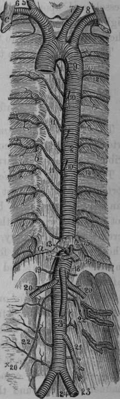
Fig. 34. The. Aorta.
1, Arch of the Aorta. 2, Thoracic Aorta. 3, Abdominal Aorta. 4, Innominate Artery. 5, Right common Carotid. 6, Right Subclavian. 7, Left common Carotid. 8, Left Subclavian. 9, Bronchial Artery, a small branch of the Aorta, 10, CEsophageal Arteries. 11, Intercostal Arteries of the right side. 12, Of the left side. 13, Phrenic Arteries. 14, Coeliac Axis. 15, Coronary Artery. 16, Splenic Artery. 17, Hepatic Artery. 18, Superior Mesenteric Artery. 19, Suprarenal Arteries. 20, Spermatic Arteries. 21, inferior Mesenteric Artery. 22, Lumbar Arteries. 23, Common Iliac Arteries. 24, Middle Sacral Artery, a, Aortic Orifice of the Diaphragm, b, Articulation of the head of the ribs, c, Anterior.
The branches of the thoracic aorta are the following:
Pericardial. (Esophageal.
Bronchial. Posterior Mediastinal.
Inferior Intercostal.
The Pericardial Branches
The Pericardial Branches are a few small and irregular arteries which arise from the front of the vessel and are distributed to the back part of the pericardium.
Continue to: