The Middle Portion Of The Arch
Description
This section is from the book "Anatomy Of The Arteries Of The Human Body", by John Hatch Power. Also available from Amazon: Anatomy of the Arteries of the Human Body, with the Descriptive Anatomy of the Heart.
The Middle Portion Of The Arch
The Middle Portion Of The Arch passes obliquely upwards, backwards, and to the left side, so that the term transverse, usually applied to it, is not correct: it terminates on the left side of the body of the second dorsal vertebra: posteriorly it is related to the lower extremity of the trachea, to the great cardiac plexus of nerves, to the thoracic duct, and left recurrent nerve : anteriorly, to the thymus gland in the early periods of life; to the left pneumogastric nerve, and also to the recurrent nerve, and to some small branches of the sympathetic nerve, derived from the superior cardiac nerve, which here unite with the recurrent: above it are the left vena innominata, to which it is united by a dense aponeurosis, connected below with an expansion of the fibrous layer of the pericardium, and above with a deep-seated process of the cervical aponeurosis, which covers the origins of the carotid and subclavian arteries, and the arteria innominata : the origins of the great arterial trunks given off regularly from this stage of the aorta, viz., the arteria innominata, the left carotid, and left subclavian, are necessarily situated above it. Beneath it, or corresponding to its concave portion, are, the left recurrent nerve, the right pulmonary artery, portion of the left auricle, the root of the left lung, sometimes the cardiac ganglion of Wrisberg, and the ligamentous chord which in intrauterine life had been the ductus arteriosus: this structure enters the concavity of the arch at a point corresponding inferiorly to the origin of the left subclavian artery from the convexity of the vessel, but a little nearer to its left side. The left recurrent nerve curves underneath that portion of the aorta which is joined by the ductus arteriosus, so that the nerve embraces within its curve the termination of the ligamentous remains of this latter vessel as well as the concavity of the arch of the aorta.
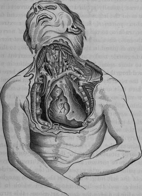
Fig. 6. Dissection to show the relations of the Vessels and Nerves in the lower part of Neck; and some of the relations of the Arch of the Aorta and its Branches. Pericardium opened, and portions of Heart exposed.
A, Right Ventricle of the Heart. B, Pulmonary Artery and Infundibulum. C, Ascending Aorta, D, Right Auricular Appendix. E, Pericardium. F, Superior Vena Cava. G, Left Pneumogastric Nerve, with loop of left Recurrent Nerve: Phrenic Nerve to their left side. H, Middle portion of the Arch of Aorta. I, Left Vena Innominata. K. Right Vena Innominata. L, Lower end of left Internal Jugular Vein cut. M, Right Subclavian Vein, N, Right Internal Jugular Vein about to join Subclavian Vein. O, Arteria Innominata. P, Right Subclavian Artery crossed by Right Pneumogastric Nerve, and in loop of Right Recurrent Nerve. Q, Right common Carotid Artery. R, Left common Carotid Artery. S, Left Subclavian Artery in relation with left Pneumogastric Nerve. T, Third stage of left Subclavian Artery. V, Left Scalenus Anticus Muscle, with Phrenic Nerve. W, W, First Ribs. X, X, Fifth Ribs cut across. Y, Y, Right and Left Mamillae. Z, Lower part of Sternum, a, Thyroid body, b, Trachea, c, Left Interual Jugular Vein cut across, d, Left Subclavian Vein, e, Clavicle cut across, and drawn downwards, f, Brachial Plexus of Nerves, g. Inferior Thyroid Artery, passing behind the cut extremity of Internal Jugular Vein, Pneumogastric Nerve, and Carotid Artery.
Continue to:
- prev: The Anterior Or Ascending Portion
- Table of Contents
- next: The Posterior Or Descending Portion Of The Arch