The Splenic Artery
Description
This section is from the book "Anatomy Of The Arteries Of The Human Body", by John Hatch Power. Also available from Amazon: Anatomy of the Arteries of the Human Body, with the Descriptive Anatomy of the Heart.
The Splenic Artery
The Splenic Artery proceeds from its origin to the left side in a very tortuous manner along a groove in the back part of the upper margin of the pancreas: posterior to it are the left crus of the diaphragm, left semilunar ganglion, and supra-renal capsule of the same side; the stomach covers it in front, and the splenic vein lies inferior to it, and in the same groove in the pancreas. Whilst the artery is remarkable for its tortuosity, the vein presents a comparatively straight course from the hilus lienis to its termination in the porta. On approaching the spleen, the artery divides into five or six terminating branches, which enter the fissure, or hilus lienis, on its concave surface. The branches given off by the splenic artery in this course are, first, small branches, pancreaticae parvae, variable in number, to the pancreas : secondly, a large branch to the pancreas, pancreatica magna, which sometimes accompanies the duct of this gland, but is often deficient: thirdly, the tasa brevia, some of which come from the trunk of the splenic, and others from the branches which enter the spleen; they are five or six in number, and are reflected to the bulging extremity of the stomach, where they communicate freely with the other arteries supplying this organ : lastly, the gastro-epiploica sinistra, which sometimes arises from one of the terminating branches of the splenic, and proceeds from left to right along the great curvature of the stomach, to anastomose with the gastro-epiploica dextra, and to give off similar ascending and descending branches.
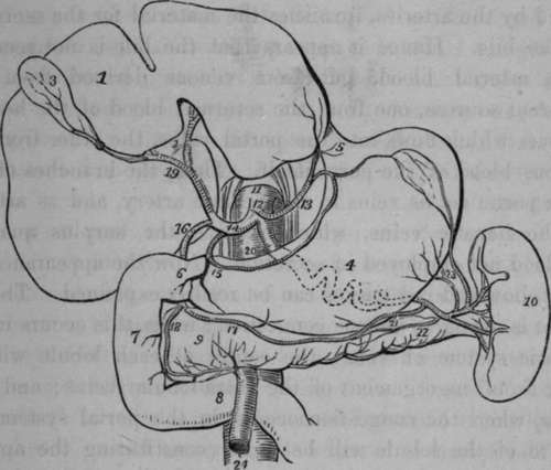
Fig. 36. Distribution of the Cceliac Artery.
1, Liver turned upward, and showing its lower surface. 2, Transverse Fissure. 3, Gall-bladder. 4, Stomach. 5, CEsophagus. 6, 7, 8, Duodenum. 9, Paucreas. 10, Spleen. 11, Aorta. 12, Coeliac Artery. 13, Coronary Artery. 14, Hepatic Artery. 15. Pyloric Artery. 16, Gastro-duodenal Artery. 17, Right Gastroepiploic Artery. 18, Pancreaticoduodenal Artery. 19, Hepatic Artery dividing into the right and left branches for the Liver. 20, Splenic Artery ; its course indicated behind the stomach by dotted lines 21, Left Gastro-epiploic Artery. 22, Pancreatic branch. 23, Gastric branches. 24, Superior Mesenteric Artery, emerging from between the Pancreas and Duodenum.
When the arteries penetrate the spleen, they soon break abruptly into numerous fine branches, compared to the hairs of a camel-hair pencil: these branches do not communicate with each other, but terminate in veins that form numerous plexuses. There are no cells in the spleen, as formerly represented. The following passage, describing the structure of the spleen, is extracted from Baly's translation of Muller:
" The spleen is invested by a strong fibrous membrane, which sends numerous band-like processes into its interior, so as to support the soft, pulpy, red tissue of the organ. In the red substance there are contained, in many animals, whitish, round corpuscles, visible by the naked eye, which were first discovered by Malpighi, and of which the existence in the human spleen has been at one time admitted, at another denied.
" The red pulpy substance consists of *a mass of red-brown granules, as large as the red particles of the blood, but differing from them in form, being irregularly globular, not flattened. These granules are easily separable from each other. In the mass which they form, the minute arteries ramify in tufts, and terminate in the plexus of venous canals, into which all the blood of the spleen is poured before it is carried out of the organ by the splenic vein. The anastomosing venous radicles, which are of considerable size, appear to have scarcely any distinct coats. If a portion of the pulpy substance of the spleen is examined more closely, it is seen to be everywhere perforated with small foramina, which are spaces bounded by the reticulated substance of the organ. These spaces are venous canals: on inflating them the organ acquires a cellular appearance; and if they are injected with wax, the substance of the spleen will present a great resemblance to the corpora cavernosa penis. There are no true cells in the spleen. The white corpuscles are imbedded in the pulpy substance, and not contained in cells, as Malpighi supposed. A fibrous trabecular tissue intersects in all directions the very soft, pulpy red substance, and affords support to the texture of the organ".
Cruveilhier observes, that if we inject the arteries of the spleen, it enlarges slowly; but if we inject the veins, it enlarges at once, showing that the connection between the arteries and the venous plexus is not so free as between the latter and the veins.
In speaking of the veins of the spleen, Mr. Gray mentions three modes in which these vessels commencefirst, as continuations of the capillaries of the arteries; this is the most common method : secondly, in intercellular spaces in the substance of the pulpy material of the spleen, through which the veins communicate with each other: thirdly, by forming an imperfect capsule to each Malpighian corpuscle. This last mode of the commencement of the veins of the spleen has not been described by other writers : Mr. Gray considers that the secretion of the Malpighian corpuscles is carried into the circulation through these small veins which form this capsule.
Continue to: