Operation Of Tying The Glutaeal Artery
Description
This section is from the book "Anatomy Of The Arteries Of The Human Body", by John Hatch Power. Also available from Amazon: Anatomy of the Arteries of the Human Body, with the Descriptive Anatomy of the Heart.
Operation Of Tying The Glutaeal Artery
M. Lizars gives the following rule for finding the trunk of the glutaeal artery. Draw a line from the posterior superior spinous process of the ilium downwards to the mid-point between the tuberosity of the ischium and the great trochanter; and then divide this line into three equal parts; the glutaeal artery will be found emerging from the pelvis at the junction of its upper and middle thirds. It will rarely be necessary, however, to apply this rule, unless for the purpose of avoiding it in opening deep abscesses of the glutaeal region; for in case of a wound, we must be guided by the wound itself; and, in case of glutaeal aneurism, the surgeon may prefer tying the internal iliac artery. The opposite practice has, no doubt, been successful: thus, Mr. Bell cut down on the tumor, in a case of glutaeal aneurism, opened the sac, and tied the vessel successfully.* Mr. Carmichael tied the glutaeal artery for a wound of this vessel by a pen-knife. The following is Mr. Carmichael's description of the operations:"The patient being placed upon a table, lying on his face, I commenced by making an incision five inches in length, beginning an inch below the superior posterior spinous process of the ilium, and about the same distance from the margin of the sacrum, and continued it in a line extending obliquely downwards to the trochanter major. The glutaeus maximus and medius were then rapidly divided, or rather their fibres separated (as the incision ran in the direction of the fibres), to the same extent as that of the integuments. The coagulated blood forming the tumor then became apparent through the sac or condensed cellular membrane with which it was covered. This was divided the whole extent of the incision by running a buttoned bistoury quickly along the finger introduced into the sac, and its contents, consisting of from one to two pounds of coagulated blood, were emptied rapidly out with both hands into a soup-plate, which it completely filled. A large jet of fresh blood instantly filled the cavity I had emptied; but, the precise spot from whence it came being perceived, I was enabled, by pressure with the finger, to prevent any further effusion, while that which had been just poured out was removed by the sponge. It was obviously the trunk of the glutaeal artery, just as it debouches from the ischiatic notch, which had been wounded. I endeavored, but in vain, to secure the artery by means of a tenaculum. I had then recourse to a common needle of large size, and with this instrument was immediately successful in passing a ligature around the bleeding vessel, and in preventing all further hemorrhage. The ligature came away on the sixth day, and the patient recovered."*
* Principles of Surgery, vol. i. p. 421.
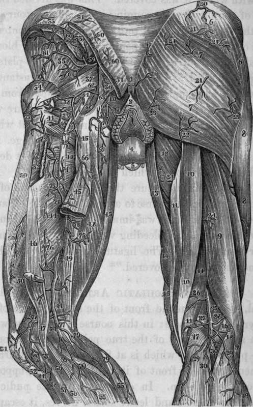
Fig. 45. Represents the Arteries of the posterior part of the Pelvis and Thigh.
1, The Coccyx. 2, The Superficial Sphincter of the Anus. 3, The Anus. 4, The Scrotum. 5, The Glans Penis. 6,6, The Glutaeus Medius Muscle. 7, 7, The Glutaeus Maximum. 8, 8, External portion of Vastus Externus. 9, 9, Biceps. 10, 10, The Semi-tendinosus. 11, 11, 11, The Semimembranosus. 12. The Adductor Magnus. 13, The Gracilis. 14, The Sartorius. 15, Small portion of the Vastus Internus. 16, The Plantaris. 17, 18, The two heads of the Gastrocnemius. 19, The Soleus. 20, Branch from the Iliolumbar Artery. 21, 21, 21. 21, Branches of the Gluteal Artery. 22, 22, Twigs from the Sciatic Artery. 23, Twig from the Internal Pudic Artery. 24,24, Branches of the Perforating Arteries. 25, The Popliteal Artery. 26, Muscular Branch from the Popliteal Artery. 27, Superior Internal Articular Artery. 28, Superior External Articular Artery. 29 . 29, Sural Arteries, proceeding from a common trunk. Upper30, Twig to Plantaris. Lower30, Branch to accompany the Posterior Saphena Vein. 31, 31, Origin of the Glutaeus Mnximus, cut. 32, Insertion of the Glutaeus Maximus, cut. 33, 33, Origin of the Glutaeus Medius, cut. 34, The insertion of the Glutaeus Medius, cut. 35, The Glutaeus Minimus. 36, The Great Sacro-Sciatic Ligament. 37, The Pyriformis. 38, 38, 39, The two Gemelli, and Obturator Internus between. 40, Portion of Levator Ani. 41, Quadratus Femoris. 42, Great Sciatic Nerve, cut. 43, Gracilis. 44, 44, The Adductor Magnus. 45, 45, 45, Long portion of Biceps Muscle, cut. 46, Short portion of Biceps between the Vastus Externus and the Adductors. 47, Tendon of Biceps. 48, The Semi-tendinosus. 49, The Semimembranosus. 50, 50, 50, Vastus Ex.
Continue to: