The Popliteal Artery
Description
This section is from the book "Anatomy Of The Arteries Of The Human Body", by John Hatch Power. Also available from Amazon: Anatomy of the Arteries of the Human Body, with the Descriptive Anatomy of the Heart.
The Popliteal Artery
This artery extends from its entrance into the popliteal space, through the opening already described, to the lower margin of the popliteus muscle. Situated at first behind the femur above its internal condyle, it runs obliquely downwards and outwards, and terminates inferiorly, corresponding to the middle line of the limb. Its anterior surface corresponds superiorly to the posterior surface of the femur; lower down, to the ligamcntum posticum of Winslowe, from which it is separated by one or two lymphatic glands; and still lower down, to the fleshy fibres of the popliteus muscle Throughout its extent its posterior surface is covered by the skin and superficial fascia, and by the popliteal fascia, together with a considerable quantity of adipose and areolar tissue : in the upper part of the space it is covered superiorly by the semi-membranosus muscle; in the middle of its course it is covered by its own vein and by the popliteal nerve, frequently by a lymphatic gland, and inferiorly by the internal head of the gastrocnemius muscle. Its vein adheres firmly to its posterior surface, projecting a little to its external side above, but to its internal side inferiorly: the popliteal nerve is much more superficial, and some adipose tissue is interposed between it and the vessels: in the superior part of this space the nerve is found at the external margin of the semi-membranosus muscle, and therefore external to the artery, while inferiorly, on account of the oblique direction of the artery, the nerve is on a plane internal to it.
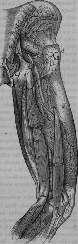
Fig. 63. Arteries of the Back of the Thigh.
1, Glutaeal Artery. 2, 3, Its superficial and deep Branch. 4, Internal Pudic Artery. 5, Ischiatic Artery. 6, Branch of the External Circumflex. 7, 8, Terminal Branches of the Perforating Arteries. 9, Popliteal Artery. 10, 11, Superior Internal and External Articular Arteries. 12, 13, Inferior Internal and External Articular Arteries. 14, Middle Articular Artery. 15, Gastrocnemial branches. a, Origin and Insertion of the Great Glutaeal Muscle, b, Origin of the Middle Glutaeal Muscle. c. Small Gluteal Muscle, d, Great Trochanter, e, Pyriform Muscle. f, Sacro-sciatic Ligaments, g, Internal Obturator Muscle, h. Quadrate Femoral Muscle, i, Sciatic Nerve, j. Tuberosity of the Ischium, k. External Vastus Muscle. l, Great Adductor, m, Short Head of the Biceps, n, Long Head, o, p, Semimembranous and Semi-tendinous Muscles. q, Gracilis, r, Gastrocnemius.
The student would do well to attend again to the relative positions of the popliteal nerve and vessels : at the upper part of the space, and passing from without inwards, he will find, first the nerve, then the vein, and more internally the artery; about the centre of the space, that is, between the two condyles, they are grouped together, and do not lie obliquely with regard to each other, but, passing from behind forwards, the nerve is most superficial, the vein lies in front of it, and still deeper and nearer to the bone we find the artery. At the lower part of the space these parts are again placed obliquely with regard to one another,the nerve is found most internally, the vein comes next, and lastly, most externally, we find the artery. Notwithstanding these alterations, throughout the entire of the space the nerve lies nearest to the skin, the artery nearest to the bone, and the vein corresponding to a plane between them both.
Continue to: