The Internal Iliac Artery
Description
This section is from the book "Anatomy Of The Arteries Of The Human Body", by John Hatch Power. Also available from Amazon: Anatomy of the Arteries of the Human Body, with the Descriptive Anatomy of the Heart.
The Internal Iliac Artery
This artery arises from the common iliac on a plane posterior to the origin of the external iliac; it is from an inch and a half to two inches in length: in the adult, it descends backwards and inwards in front of the sacro-iliac symphysis, as far as the superior extremity of the great sacro-sciatic notch ; here it becomes ligamentous, and ascends to the umbilicus, on the side of the bladder, being covered posteriorly by its superior false ligament. In its first or truly arterial stage it forms a curvature, the concavity of which looks forwards. Its posterior or convex surface rests on the sacroiliac symphysis, from which it is separated by its corresponding vein, which on the right side projects from underneath its outer edge; and by the lumbo-sacral nerve, which lies still deeper than the vein: from behind it we see also the obturator nerve passing forwards, and running in the angle between the internal and external iliac arteries. Its anterior surface is covered by peritoneum, and crossed superiorly, at its origin by the ureter, and inferiorly by the vas deferens. In addition to these, the rectum covers the artery on the left side, and the bladder forms an anterior relation to the internal iliac arteries of both sides.
The Internal Iliac Artery Of The Foetus
The Internal Iliac Artery Of The Foetus presents for our consideration many distinct peculiarities. First, it is considerably larger than the external iliac; the reverse is the fact in the adult: in the foetus, it does not descend deep into the pelvis, but rather winds along the ilio-pectineal line, and then ascends, not in a ligamentous form, but pervious, and carrying blood from the foetus along the sides of the bladder through the umbilicus to the placenta. From the umbilicus to the placenta the two arteries form part of the umbilical cord.
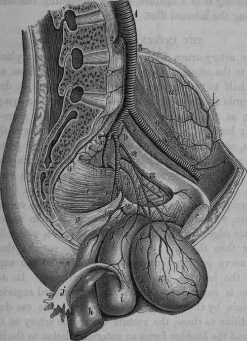
Fig. 44. View of the left side of the Pelvis, the Bladder, Uterus, Vagina, and Rectum, turned downward so as to exhibit the distribution of the Internal Iliac Artery.
1, Aorta. 2, Right Common Iliac Artery. 3, Left Common Iliac, 4, Middle Sacral. 5, External Iliac. 6, Circumflex Iliac. 7, Epigastric. 8, Internal Iliac. 9, Ilio-Lumbar. 10, Lateral Sacral Arteries. 11, Glutaeal Artery passing from the Pelvis, above the Pyriform Muscle, at the upper part of the great Sacro-Sciatic Foramen. 12, Superior Vesical Artery ; the branch cut off is extended into the remains of the Umbilical Artery. 13, Obturator Artery. 14, Inferior Vesical Artery giving off the Uterine Artery to the Vagina and Uterus. 15, Middle Haemorrhoidal Artery. 16, Internal Pudic Artery, seen emerging from and again entering the Pelvis. 17, Ischiatic Artery, a, Iliac Muscle. 6, Psoas Muscle, c, Symphysis of the Pubis, d, Sacrum, c, Pyriform Muscle. f, Internal Obturator Muscle. g, Sacro-Sciatic Ligaments, ft, Rectum, i. Uterus and Vagina, j. Fallopian Tube. Bladder.
After birth, the internal iliac arteries gradually diminish, and the external iliac arteries, and posterior or external branches of the internal iliac, gradually enlarge.
Continue to: