The Liver
Description
This section is from the book "Landmarks And Surface Markings Of The Human Body", by Louis Bathe Rawling. Also available from Amazon: Landmarks and Surface Markings of the Human Body.
The Liver
The anterior border can be mapped out by drawing a curved line from a point in the fifth left interspace 3 1/2 inches from the middle line (the position of the apex of the heart), (Fig. XX, 2.) the line cutting the left costal margin at the tip of the eighth costal cartilage and the right costal margin at the tip of the ninth costal cartilage. Between these two latter points, the anterior border of the liver crosses the middle line half-way between the umbilicus and the sterno-xiphoid junction (= transpyloric plane), whilst a notch to the right of the middle line indicates the hepatic attachment of the ligamentum teres, (Fig. XX, 4.) which passes from that notch downwards and inwards to the umbilicus.
Beyond the tip of the ninth right costal cartilage the anterior border of the liver follows the lower limit of the costal arch, descending sometimes even below that level, (Fig. XVII, 12.) and after cutting across the twelfth rib, ascends towards the level of the eleventh dorsal spine. The upper limit of the liver is indicated by a line starting as before in the fifth left interspace 3 1/2 inches from the middle line, Fig. XX. and ascending slightly as it passes to the right. This line cuts across the sixth right chondro-sternal articulation, (Fig. XVII, 12.) the upper border of the right fifth costal cartilage in the right lateral vertical plane, the sixth rib in the mid-axillary line, sweeping thence just below the angle of the scapula towards the eighth dorsal spine.
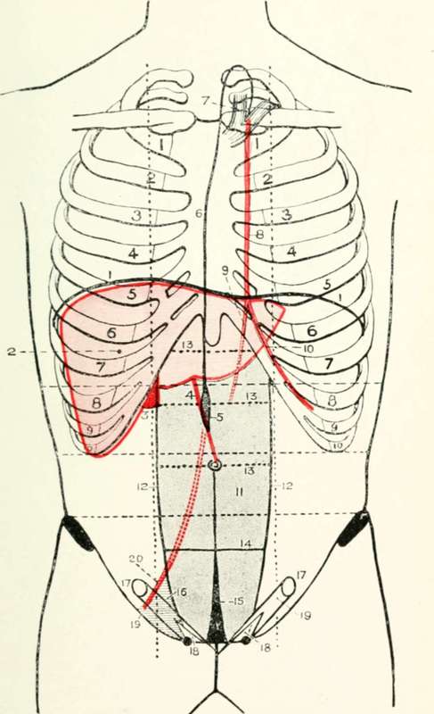
Fig. XX. The Liver, Anterior Abdominal Wall, Etc
1. The diaphragm.
2. The liver.
3. The gall-bladder.
4. The ligamentum teres.
5. The receptaculum chyli.
6. The thoracic duct.
7. The venous termination of the duct
8. The internal mammary artery.
9. The superior epigastric artery.
10. The musculo-phrenic artery.
11. The rectus abdominis muscle.
12. The linae semilunares. l3. The linae transversae.
14. The semilunar fold of Douglas.
15. The urachus.
16. Hesselbach's triangle.
17. The internal abdominal ring.
18. The external abdominal ring.
19. The inguinal canal.
20. The deep epigastric artery,
The Gall-Bladder
(Fig. XX, 3.) The fundus projects from under the anterior border of the liver in the angle between the tips of the ninth and tenth costal cartilages and the outer border of the rectus abdominis muscle. The diaphragm. (Fig. XX, 1.) On ordinary inspiration the right dome of the diaphragm corresponds in level to the lower part of the fourth right interspace, whilst the left dome ascends to the lower part of the fifth left rib and costal cartilage.
Continue to: