The Brachial Plexus
Description
This section is from the book "Landmarks And Surface Markings Of The Human Body", by Louis Bathe Rawling. Also available from Amazon: Landmarks and Surface Markings of the Human Body.
The Brachial Plexus
The upper limit of the nerve-(Fig. III, 17., Fig. IV, 7.) trunks which form this plexus is represented by a line drawn from the mid-point between the anterior and posterior borders of the sterno-mastoid muscle at the level of the cricoid cartilage to a second point situated just external to the mid-point of the clavicle. The lowest cord lies behind the third part of the subclavian artery.
The rima glottidis, which in its front part is laterally bounded by the true vocal cords, lies opposite Fig. V, 10. the mid-point along the anterior border of the thyroid cartilage.
The epiglottis, though fixed below to the thyroid angle immediately above the point of attachment of Fig. V, 9. true vocal cords, extends upwards to above the level of the body of the hyoid bone.
A suicidal cut-throat frequently involves the thyro-hyoid space, and the epiglottis may be severed from its thyroid attachment.
The isthmus of the thyroid gland crosses the trachea Fig. V, 12. about 1/2 to 3/4 inch below the cricoid cartilage. Fig. V. The structures in the middle line of the neck :
(1) Passing down from the jaw to the body of the hyoid bone, the two genio-hyoid muscles lie each side of the middle line. They are placed, however, deep to the mylohyoid muscles, which are directed downwards and inwards to the median raphe and to the body of the hyoid bone. (2) The body of the hyoid bone.
(3) The thyro-hyoid space.
(4) The thyroid cartilage.
(5) The crico-thyroid space.
(6) The cricoid cartilage.
(7) The upper two or three tracheal rings.
(8) The isthmus of the thyroid gland (9) The trachea.
(10) The suprasternal notch.
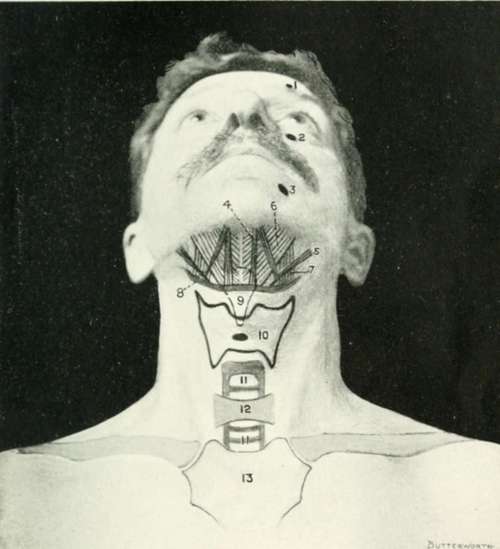
Fig. V. The Front Of The Neck
1. The supra-orbital foramen.
2. The infra-orbital foramen.
3. The mental foramen.
4. The genio-hvoid muscle (deep to the mylo-hyoid muscle).
5. The digastric muscle.
6. The mylo-hvoid muscle.
7. The hyo-glossus muscle.
8. The hvoid bone.
9. The epiglottis.
10. The thyroid cartilage.
11. 11. The trachea.
12. The thyroid isthmus.
13. The sternum.
Continue to: