Chapter IV. The Abdomen
Description
This section is from the book "Landmarks And Surface Markings Of The Human Body", by Louis Bathe Rawling. Also available from Amazon: Landmarks and Surface Markings of the Human Body.
Chapter IV. The Abdomen
The anterior aspect of the trunk (i.e; thorax and abdomen, Fig. XVIII,3.) is divisible into right and left halves by a median vertical plane from the middle point at the suprasternal notch above to the symphysis pubis below.
Each half is again divided by a lateral vertical plane which is drawn parallel to the median plane, (Fig. XVIII, 4.) half-way between that plane and the anterior superior iliac spine. Prolonged downwards, this lateral plane crosses Poupart's ligament rather nearer to the inner than to the outer end. Prolonged upwards, it crosses the clavicle about midway between the median point at the suprasternal notch and the acromio-clavicular joint.
That part of the lateral vertical plane which traverses the mammary region is sometimes called the "mammary plane," that part which crosses the clavicle the "clavicular plane," and the downward prolongation which cuts across Poupart's ligament the "Poupart plane."
The clavicular, mammary, and Poupart planes are, however, continuous, and they together form the lateral vertical plane, which is chosen in preference to the mid-Poupart plane of many anatomists, since it is measured from the median plane to a fixed bony point. Two vertical planes only will be consequently retained in the subdivision of the anterior aspect of the trunk, the median and lateral vertical planes.
The median plane can be bisected by a horizontal plane which is on the same level as the body of the first lumbar vertebra. This plane so constantly cuts across the pyloric end of the stomach that it is called the transpyloric plane, and it will be found that not only does this plane lie half-way between the suprasternal notch and the symphysis pubis, (Fig. XVIII.) but that it also lies midway between the umbilicus and the sterno-xiphoid junction. It is, therefore, not necessary to expose the whole of the anterior aspect of the trunk in order to verify the position of the transpyloric plane, a plane of the greatest value in defining the position of several abdominal viscera.*
The point at which the median vertical and transpyloric planes intersect has been suitably called the "central point," (Fig. XVIII, 1.)and the point of intersection of the lateral vertical and transpyloric planes may be tentatively called the "lateral central" or " paracentral " point. (Fig. XVIII, 2.)
This latter point usually corresponds to the anterior extremity of the ninth costal cartilage.
The distance between the "central point" and the top of the symphysis pubis is bisected by a horizontal plane which passes through the tubercles of the iliac crests, (Fig. XVIII.) the intertubercular plane, a plane corresponding to the level of the body of the fifth lumbar vertebra. It has also been suggested that the distance between the "central point" and the suprasternal notch should likewise be bisected by a plane, the thoracic plane, which crosses the gladiolus at the level of some part of the anterior extremities of the fourth costal cartilages.
* N.B
Most of the " planes " mentioned in the text were first put forward by Dr. C. Addison, Lecturer and Senior Demonstrator of Anatomy, St. Bartholomew's Hospital, to whom the credit is due.
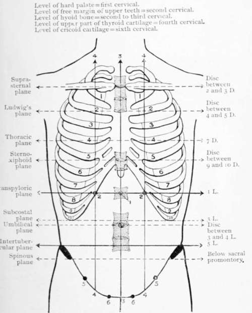
Fig. XVIII. The Abdominal And Thoracic Planes
1. The central point.
2. The lateral central or paracentral point.
3. The median vertical plane.
4. The lateral vertical plane.
5. The mid-Poupart point.
6. The pubic spines.
This plane is, however, of little value, (Fig. XVIII.) and is merely mentioned as completing the symmetrical subdivision of the median vertical plane into four equal parts.
The following planes are, therefore, chosen as the most scientific in the subdivision of the anterior aspect of the trunk :
Two vertical planes(1) The median; (2) the lateral.
Three transverse planes(1) The intertubercular; (2) the transpyloric ; (3) the thoracic.
Two important points are also named(1) The central point; (2) the lateral central point.
The abdominal regions mapped out by the intersection of the transpyloric and intertubercular planes with the lateral vertical planes receive the same nomenclature as in the older methods of regional subdivision of the abdomen. These regions are nine in number: Fig. XVIII.
1. Right hypochondriac.
2. Epigastric.
3. Left hypochondriac.
4. Right lumbar.
5. Umbilical.
6. Left lumbar.
7. Right iliac.
8. Hypogastric.
9. Left iliac.
Continue to:
- prev: The Trachea And Bronchi
- Table of Contents
- next: Other Transverse Planes, With Their Corresponding Vertebral Levels