67. The Mucous Membrane Of The Stomach
Description
This section is from the book "Animal Physiology: The Structure And Functions Of The Human Body", by John Cleland. Also available from Amazon: Animal Physiology, the Structure and Functions of the Human Body.
67. The Mucous Membrane Of The Stomach
The Mucous Membrane Of The Stomach, by which the gastric juice is secreted, is thick, soft, and smooth. Looked at under the microscope, the surface is seen to be thrown into shallow pits, into each of which open several small glands; and in sections vertical to the surface the mucous membrane is shown to consist in greater part of tubular glands, sometimes branched, called the gastric follicles, which may reach 1/20 inch in length, and are crowded together. They are all lined with columnar epithelium down to the bottom, and the epithelial cells have been seen to undergo a change of appearance in digestion, when the gastric juice is secreted; but the majority of the follicles likewise contain numbers of large oval and more easily discovered cells underneath the columnar epithelium, structures of uncertain function, which, from having been supposed to be the agents in secreting pepsin, have been named peptic cells.
The glands in which these cells occur are called peptic glands, while the others are termed mucous. In some animals, for example the dog, certain regions of the stomach contain none but peptic glands, while the pyloric region has none but mucous glands; and this circumstance has been taken advantage of to determine the character of the secretion furnished by the two kinds of glands. But while one observer (Ebstein) gives a most careful and convincing account of experiments to prove that both kinds secrete pepsin, numerous others come to a different conclusion ; and it may be considered as still undecided whether the peptic or the columnar cells are the agents by which pepsin is really secreted.
68. Immediately beyond the pyloric valve, on entering the intestine, the mucous membrane changes its character. Not only is it firmer and thinner, but it is covered over with minute thread-like projections like velvet pile, named villi, best brought into view by laying the opened intestine in water, so as to make them float. They are as much as half a line long in the duodenum, and extremely crowded, but gradually get shorter and fewer in the lower parts of the small intestine, until, near the termination of the ileum, they degenerate into scattered wartlike eminences. The villi are confined entirely to the small intestine, and are a means of increasing the extent of mucous membrane for the purpose of absorption; each villus being dipped like a finger in the chyme, and sucking up the digested parts of it by every portion of its surface. The absorbent surface of the small intestine is further increased by crescentic folds of the mucous membrane passing more than half round it, and dipping into the interior. They are called valvulae conniventes, and, like the villi, are most numerous and well developed in the duodenum and jejunum.
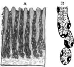
Fig. 54. Gastric Follicles. A, Vertical section of mucous membrane from the middle of the human stomach. B, A more highly magnified view of portion of a gastric follicle of a dog, showing both peptic and columnar cells. From Heidenhain.
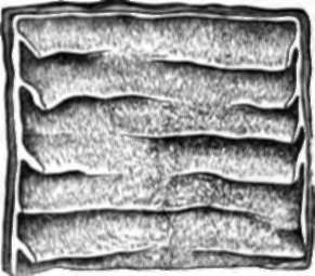
Fig. 65. Valvule Conniventes, exhibited in a portion of jejunum cut open. Two-thirds natural size.
The thickness of the mucous membrane of the small intestine is occupied, like that of the stomach, with closely set tubules, only they are much smaller than those of the stomach, contain no other than columnar epithelium, and are quite simple; these are called the follicles or crypts of Lieberkuhn. The duodenum has likewise three other secretions poured into it, namely, the bile, the pancreatic juice, and the secretion of a number of minute glands of a lobu-lated description, scattered beneath its mucous membrane, and called Brunner's glands.
Continue to: