115. The Absorbents
Description
This section is from the book "Animal Physiology: The Structure And Functions Of The Human Body", by John Cleland. Also available from Amazon: Animal Physiology, the Structure and Functions of the Human Body.
115. The Absorbents
The Absorbents which come from the small intestine, although they in no way differ from the "lymphatics of the rest of the body, are distinguished as lacteals. The distinction is unscientific, inasmuch as the lacteals are simply the lymphatics of the small intestine, carrying lymph and nothing else when the intestine is empty; but the name arose naturally enough from the milky appearance of their contents during digestion, and must be submitted to. The lacteals pass from the intestine back between the folds of the mesentery to reach the receptaculum chyli, and in their course traverse a plentiful group of lymphatic glands, named, from their position, mesenteric, and subject, like other lymphatic glands, to be irritated into inflammation and disease, when the fluids reaching them by their afferent vessels are altered by inflammation of the tissues from which they are derived. Moreover, such disease, unfortunately common in weak children, is of much graver importance in the instance of these than of other lymphatic glands, since it interferes with the passage of nourishment from the intestine into the blood. This constitutes the essence of the disease called tabes mesenterica.
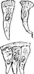
Fig. 82. Colum-narEpithelial Cells op Intestine, highly magnified.show-ing the striated substance on their free extremities.
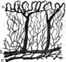
Fig. 83. Blood-vessels op Mucous Membrane of Stomach : vertical section magnified, a, Venous radicles descending from the free surface ; 5, veins ; c, arteries.
Every villus presents on its surface a coat of columnar epithelium continuous with that of the rest of the mucous membrane, and beneath this a network of capillary blood-vessels, while in the centre there is a lacteal. In the lower animals there are sometimes several lacteals forming loops in one villus; but in the human subject there is usually only one in each. This lacteal is somewhat dilated at its extremity, and it does not appear at that part to have any special wall.
When absorption is going on, the epithelial cells of the villi become turbid with molecules of oil, the tissue beneath becomes turbid likewise, and from the tissue the lacteals are filled. The free extremity of each columnar epithelial cell presents a thick layer of substance less consistent than the rest of the cell wall, but more consistent than its contents, and sometimes having a vertically streaked appearance. Through this substance the molecules of emulsified oil find their way; and afterwards they pass onwards through fine prolongations of the deep ends of the epithelial cells : these appear to communicate with branches of connective-tissue-corpuscles, by which in like manner the molecules are carried to the lacteal.
Only a small quantity of oil is taken into the capillary bloodvessels, by far the greater amount being absorbed by the lacteals; and this, indeed, is the most notable difference between lacteal and capillary absorption, so far as nutritive matters are concerned, for it is very certain that saccharine and albuminoid substances are taken up freely by both sets of vessels. The arrangement by which the blood-vessels are enabled to furnish material for the nourishment of texture, and formation of secretions, while they also absorb matters presented to them in both stomach and intestine, is a very beautiful one. It is plain that blood, to which variable amounts of chance ingredients had been added, would be unfit for purposes of nutrition; but the difficulty is got over in this way, that the arterioles break up into capillaries in the deep part of the mucous membrane, which first supply the glandular structure, and then open into venous radicles which take origin close to the surface; so that the blood, in its purity, nourishes the glands, and does not take up foreign matters until about to enter the veins.
Continue to: