16. The Thigh Bone Or Femur
Description
This section is from the book "Animal Physiology: The Structure And Functions Of The Human Body", by John Cleland. Also available from Amazon: Animal Physiology, the Structure and Functions of the Human Body.
16. The Thigh Bone Or Femur
The Thigh Bone Or Femur corresponds with the humerus of the upper limb; in front of the knee is the patella or kneecap, which is a sesamoid bone or ossification within a tendon, and not at all correspondent with the olecranon of the elbow: in the leg the tibia and fibula are the bones corresponding with the radius and ulna in the fore-arm; and in the foot there is a close correspondence of all the bones with those of the hand.
The parts of the foot are called the tarsus, metatarsus, and phalanges. The phalanges and metatarsal bones are arranged quite like those of the hand, the great toe being similar to the thumb in having only two phalanges. The bones behind the metatarsals are the internal, middle, and external cuneiform bones, supporting the three inner metatarsals, and the cuboid, supporting the fourth and fifth. These four bones obviously correspond with the four carpal bones of the lower range in the hand: but behind them are three others, the scaphoid, lying behind the three cuneiform bones; the calcaneum, a very large bone projecting back from the cuboid, and forming the heel; and the astragalus, resting on the calcaneum behind, pressing against the scaphoid in front, and articulating with the leg-bones above; and the dissimilarity of appearance of the tarsus as compared with the carpus, is due to the large and unequal development of these three bones. The scaphoid, however, corresponds with the bone of the same name in the hand, and the astragalus with the semilunar, while the Calcaneum represents the cuneiform and the pisiform together; and it is owing to the great development backwards of the calcaneum to form the heel, that the hollow by which tendons and other structures pass from the back of the leg to the sole of the foot is turned inwards so as to lie between the heel and inner ankle.
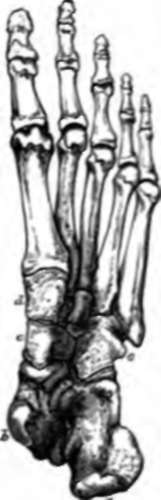
Fig. 14. Foot from below, a, calcaneum; b, astralagus; c, scaphoid; d, internal cuneiform; e, cuboid. The middle and external cuneiform are seen between the cuboid and internal cuneiform.
The same principle of conduction of pressure through tense ligaments, which we have noted in the hand and the pelvis, is resorted to again in the foot. The foot may be conveniently regarded as consisting of two arches supported behind by a common pier, the back part of the calcaneum. To the inner and principal arch belong the three inner toes, and the keystone of this is the astragalus, the fore part or 14 o head of which, lying between the calcaneum and scaphoid, is retained in position by a strong inferior calcaneo-scaphoid ligament, which has frequently to bear nearly the whole weight of the body. The outer arch is continued forwards from the calcaneum to the cuboid and two outer toes, and is prevented from falling flat by strong calcaneocuboid or plantar ligaments.
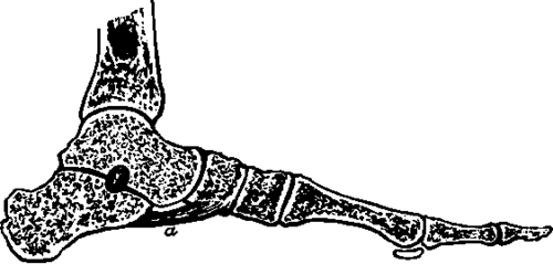
Fig. 15. Section of Foot, showing, a, the inferior calcaneoscaphoid ligament supporting the head of the astralagus.
Continue to:
- prev: 15. The Fifth Or Lowest Lumbar Vertebra
- Table of Contents
- next: 17. The First And Second Cervical Vertebræ