15. The Fifth Or Lowest Lumbar Vertebra
Description
This section is from the book "Animal Physiology: The Structure And Functions Of The Human Body", by John Cleland. Also available from Amazon: Animal Physiology, the Structure and Functions of the Human Body.
15. The Fifth Or Lowest Lumbar Vertebra
The Fifth Or Lowest Lumbar Vertebra rests on the broad upper end of a curved wedge, the sacrum, which consists of five other vertebræ fused together in one bone; and at the lower and narrow end of this bone are four more of a rudimentary description, corresponding with the caudal vertebræ or bones of the tail in other animals, but usually named by the human anatomist, collectively, the coccyx.
On its sides, in the upper two-thirds of its extent, the sacrum is closely united to the two pelvic or innominate bones, which, together with it, enclose a basin or cavity, called the pelvis. Examined in early life, each innominate bone is seen to consist of three parts, which meet at the articular cup, called the acetabulum, for the head of the thigh bone. The expanded upper part is called the ilium, the lower part is called the ischium, while the part which meets with the opposite bone in the middle line is the os pubis, and the union is called the symphysis pubis. The expanded ilium obviously corresponds with the shoulder-blade in the upper limb, and the pair of innominate bones with the shoulder-girdle, notwithstanding that the shoulder-girdle is but little connected with the trunk, while the innominate bones take an important part in bounding the visceral cavity.
From the upper end of the sacrum a prominent ring of the innominate bone can be followed round to the symphysis pubis, constituting the brim of the true pelvis as distinguished from the part of the abdomen between the blades of the iliac bones. In the erect posture of the body this ring lies at an angle of 60° with the horizontal, so that the sacrum presses downwards on it. But the sacrum is so placed that its upper end is thrown in front of the rest of it, and that its hinder surface, which is narrower than the anterior, looks upwards; it would, therefore, fall down into the pelvis were it not supported by a pair of exceedingly strong posterior sacro-iliac ligaments; and it is through these ligaments much more than by direct pressure that the weight of the body is conducted from the sacrum to the pelvis.
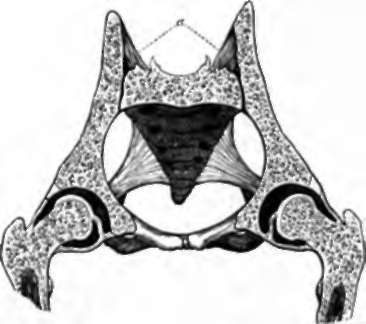
Fig. 13. Section of Pelvis, showing the suspension of the sacrum between the haunch bones, a, the posterior sacroiliac ligaments.
Continue to: