The Ilio Lumbar Artery
Description
This section is from the book "Anatomy Of The Arteries Of The Human Body", by John Hatch Power. Also available from Amazon: Anatomy of the Arteries of the Human Body, with the Descriptive Anatomy of the Heart.
The Ilio Lumbar Artery
The Ilio Lumbar Artery arises from the posterior part of the internal iliac, and takes a direction upwards, backwards, and outwards in front of the lumbo-sacral nerve, and behind the obturator nerve and psoas muscle; in this situation it divides into its two principal branches, the iliac and the lumbar.
The Iliac branch takes a transverse direction beneath the anterior crural nerve and psoas and iliacus internus muscles; some of its branches ramify on the surface of the muscle, and others in a more deep-seated situation. From the latter branches arises the nutritious artery of the ilium, which enters the canal observable near the centre of the internal iliac fossa. The Lumbar branch ascends under cover of the psoas muscle and on the front of the lumbo-sacral nerve : one of its branches enters the lateral foramen of the spine between the fifth lumbar vertebra and the sacrum, and is distributed on the tunics of the spinal marrow; the others are distributed to the psoas and quadratus lumborum muscles. This lumbar branch sometimes arises from the middle sacral artery.
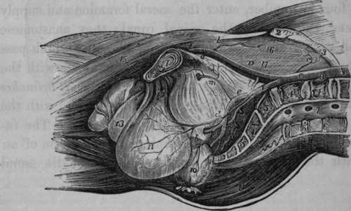
Fig. 56. Represents a lateral view of the Arteries of the Pelvis in the Male subject. A vertical incision has been carried through the Symphysis Pubis, the middle of the Lumbar Vertebrae and the Sacrum. The Viscera are drawn downwards.
A, Aorta. B, Left Common Iliac Artery divided. C, Right Common Iliac. D, Kxternal Iliac. E, Femoral Artery. F, Internal Iliac, a, Epigastric Artery cut. b, Internal Circumflexa Ilii. c, Twig from the Ilio-lumbar Artery, d, Vesical Artery, e, Anterior part of Internal Iliac Artery, f, Internal Pudic Artery. g, Sciatic Artery, h, Middle Haemorrhoidal Artery coming from the Pudic, and giving off vesical twigs, k. Posterior part of Internal Iliac Artery. 1, Iliolumbar Artery, m, Obturator Artery, n, Glutaeal Artery, o, A small branch passing into the first Sacral Foramen, p, Lateral Sacral Artery a little lower down. 1, Symphysis Pubis. 2, Anterior Superior Spine of the Ilium. 3, Crest of the Ilium. 4, 4, Divided last two Lumbar Vertebrae. 6, 5, Divided Sacrum. 6. 6, Divided spinous processes of the two last Lumbar Vertebrae. 7, Termination of the Spinal Canal. 8, Erector Spinas Muscle of the right side. 9, Glutaeus Maximus Muscle. 10, Rectum divided, tied, and turned down. 11, Bladder drawn down. 12, Anterior Ligaments of the Bladder. 13, Scrotum. 14, Corpus Cavernosum of the left side divided. 15, Sartorius Muscle. 16, Iliac and Psoas Muscle, covered by 17, the Iliac Fascia.
The communications of the ilio-lumbar artery are extremely important: its lumbar branch communicates with the proper lumbar and intercostal arteries, and its iliac branch communicates freely at the crest of the ilium with the glutaeal, circumflexa ilii, and external circumflex femoris arteries. This explains how blood is freely carried to the extremities when the iliac artery or lower part of the aorta has been rendered impervious.
Continue to: