First Stage Of The Axillary Artery
Description
This section is from the book "Anatomy Of The Arteries Of The Human Body", by John Hatch Power. Also available from Amazon: Anatomy of the Arteries of the Human Body, with the Descriptive Anatomy of the Heart.
First Stage Of The Axillary Artery
Anteriorly, it is covered by the integuments, the platysma, supra-clavicular branches of the cervical plexus, the upper portion of the pectoralis major, and immediately under cover of this muscle, by some areolar tissue, together with the expansion of fascia given off from the ligamentum bicorne; and close to the clavicle by the ligament itself, and a small portion of the inferior margin of the subclavius muscle: we find also anterior to the artery and vein the anterior thoracic nerve, small branches of which curve underneath the vessel and unite with the middle thoracic nerve which descends behind it, thus forming a nervous loop around the artery. Posteriorly, it rests against the external layer of the first intercostal muscles, and corresponds, with the interposition of some areolar tissue, to the origin which the serratus magnus takes from the second rib. Externally, it is related to the brachial plexus of nerves; these nerves lie also upon a plane somewhat above the level of the artery; the trunk formed by the union of the eighth cervical and first dorsal nerves lies nearer to the artery, and upon a plane superior, external, and posterior to this vessel. Internally, it is in close relation to the axillary vein, which, when distended with blood, overlaps the inner portion of the artery, and gets more in front of it as it descends. In this situation the vein corresponds to the two first ribs and to the upper part of the serratus magnus. Thus in the first stage the artery lies between the brachial plexus on the outside, and the axillary vein upon the inside.
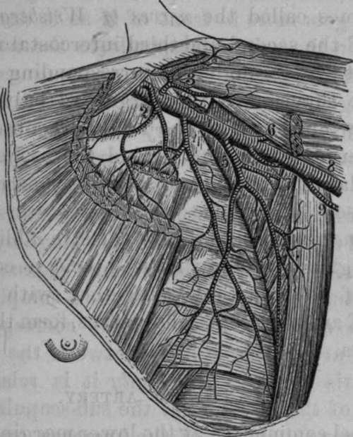
Fig. 23. View of the Axillary Artery, portions of the Pectoral and Deltoid Muscles removed.
1, Axillary Artery. 2, Superior Thoracic. 3. Acromial Thoracic. 4, Long Thoracic. 5, Subscapular. 6, Auterior Circumflex. 7, Posterior Circumflex. 8, Brachial Artery. 9, Superior Profound Artery.
Continue to: