Retina
Description
This section is from the book "Wonders Of The Human Body", by Auguste Le Pileur. Also available from Amazon: Wonders of the Human Body.
Retina
The internal surface of the choroid, or rather the pigmentary layer which covers it, is lined by the retina, a nervous membrane, upon which the objects are depicted that we see. It appears to be formed by the expansion of the optic nerve, which enters the eye at its posterior part, and forms at the bottom of the globe an enlargement, which is called the papilla of the optic nerve. The retina develops itself from the papilla, around which it forms a fold, and extends over the cavity of the eye to the circumference of the ciliary processes of the vitreous body, where, according to Cruveilhier, it abruptly terminates. It is of an opaline white colour, semi-transparent, and easily torn. Its centre, which corresponds to the antero-posterior axis of the eye, is to the outside of the papilla of the optic nerve, where there is a yellow spot (macula lutea) and a depression (fovea centralis). The yellow spot seems to be the point in the eye where vision is most distinct Microscopists describe the retina as being composed of five, or even eight layers, of which the external one is vascular, and in contact with the choroid; the internal one, very important in a physiological point of view, is the membrane of Jacob. It is composed of cylinders, or rods, joined together like the stakes of a palisade, perpendicular to the plane of the membrane, and forming by their free extremities a mosaic, each microscopic division of which is about 0.001 of a line in diameter according to Robin, and 0.0008 of a line according to Helmholtz; and represents a section of a rod. We shall see what part these terminal points play in vision.
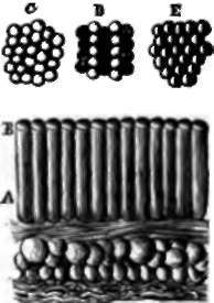
Fig. 35. Rods of Jacob under the microscope.
A. Rods of Jacob.
B. Their extremities forming surface of retina.
C. Retinal mosaic formed by the rods.
D. Points of the retinal mosaic receiving different luminous rays.
e. Points receiving each two different rays.
Continue to:
- prev: Iris
- Table of Contents
- next: Vitreous Body