The Tarsus
Description
This section is from the book "Surgical Anatomy", by John A. C. MacEwen. Also available from Amazon: Surgical Anatomy.
The Tarsus
Three sets of joints exist in the tarsus : (i) Between astragalus and os calcis (posterior). (2) (a) Between astragalus and scaphoid, (b) between os calcis and cuboid. These two together constitute the midtarsal joint. (3) (a) Between scaphoid and three cuneiforms, (b) between the cuneiforms, (c) between external cuneiform and cuboid.
1. This consists of two parts, entirely separated by the strong astragalo-calcaneal ligament which unites the two bones. The anterior portion is continuous with the joint between the head of the astragalus and the scaphoid. Comparatively weak peripheral ligaments (external, internal, and posterior) also help to unite the two bones, which are further strengthened by the lateral ligaments of the ankle and various tendons. The articulation permits of adduction with slight rotation inwards, and abduction with slight rotation outwards.
2. (a) This articulation is continuous with the last, and is of the ball and socket variety. The lower aspect of the head of the astragalus is supported by the inferior calcaneo-scaphoid ligament, which is a powerful band of triangular shape, running from the sustentaculum tali to the under surface of the scaphoid, and which again is supported on its under surface by the tendon of the tibialis posticus. There is also an external calcaneo-scaphoid and an astragalo-scaphoid ligament, the latter situated on the upper, or dorsal, surface of the articulation, (b) Each bone presents a concavo-convex surface mutually adapted to one another. The joint is supported by an internal calcaneo-cuboid ligament, which springs from the os calcis, together with an external calcaneo-scaphoid ligament, the two diverging in the form of the letter V, to be inserted into the scaphoid and cuboid respectively. There are also external and dorsal calcaneo-cuboid ligaments, while below are situated the inferior calcaneo-cuboid, or plantar, ligaments. The short plantar ligament is more deeply placed, and extends from the anterior tubercle on the inferior surface of the os calcis to the proximal part of the under surface of the cuboid. The long plantar ligament covers the short, and is separated from it by some fatty tissue. It extends from the posterior tuberosities of the os calcis along its whole inferior surface to the ridge on the under surface of the cuboid, immediately behind the peroneal groove, some fibres being continued over the groove to the bases of the second, third, and fourth metatarsals. The long plantar is a powerful ligament, and together with the short plantar and inferior cal-caneo-scaphoid ligaments plays an important part in maintaining the arch of the foot. The midtarsal joint permits of flexion and extension and rotation on an antero-posterior axis, the movement in the astragaloid section being freer than that in the calcaneal.
3. These joints are supplied with dorsal, interosseous, and strong plantar ligaments, the latter being reinforced by slips from the tibialis posticus tendon.
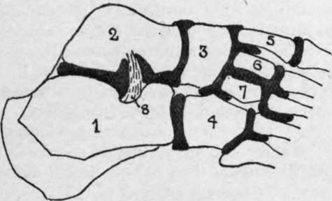
Fig. 45.-Diagram of the Six Synovial Membranes of the Foot.
1. | Os calcis. | 5 | Internal cuneiform. |
2. | Astragalus. | 6. | Middle cuneiform. |
3 | Scaphoid. | 7 | External cuneiform. |
4 | Cuboid. | 8. | Interosseous ligament. |
Continue to: