The Dorsalis Pedis
Description
This section is from the book "Surgical Anatomy", by John A. C. MacEwen. Also available from Amazon: Surgical Anatomy.
The Dorsalis Pedis
The interior tibial artery is crossed at the ankle by the tendon of the extensor hallucis, and then lies between it and that of the extensor digitorum, being continued into the foot as the Dorsalis pedis. This vessel lies in a line from the centre of the front of the ankle to the posterior extremity of the first interosseous space, being accompanied on its outer side by the anterior tibial nerve and lying between the extensor longus hallucis on its inner side, and the innermost tendon of the extensor digitorum on its outer side. The vessel, lying as it does superficially and close to the bones of the foot, is frequently divided and ruptured in wounds and injuries, and its ligature presents little difficulty, save in the cadaver, where a slightly more superficial vein is frequently mistaken for the artery.
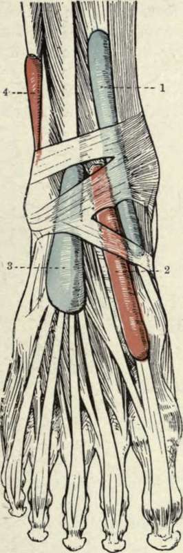
Fig. 43.-The Synovial Sheaths at the Ankle. Anterior View. (After L. Testut's " Anatomie Humaine.")
1. Tibialis amicus. | 3. Extensor longus digitorum. |
2. Extensor proprius hallucis. | 4. Peroneus longus et brevis. |
The posterior tibial artery may be ligatured at the ankle through a curved incision a finger's breadth below the internal malleolus. At the lower border of the internal annular ligament it divides into internal and external plantar arteries, and sends calcaneal branches to the heel. (The anterior and posterior branches of the peroneal artery descend to the ankle, and anastomose with the branches of the anterior and posterior tibials.) The internal plantar artery, the smaller of the two, runs forward between the abductor hallucis and flexor brevis digitorum to the head of the first metatarsal bone, while the external plantar runs forwards and outwards between the flexor brevis digitorum and abductor minimi digiti to the base of the fifth metatarsal, where it turns abruptly inwards to form the plantar arch, which crosses the bases of the metatarsals to the outer side of the base of the first metatarsal to anastomose with the dorsalis pedis. The course of the internal plantar is represented by a line from a point midway between the internal malleolus and prominence of the heel to the middle of the under surface of the great toe ; the external runs from the same point to within ½ inch of the base of the fifth metatarsal. Wounds of the plantar arch are troublesome, not only because it lies deeply and might necessitate damage to tendons and nerves to reach it, but because of the free anastomoses between the anterior and posterior tibial and peroneal arteries. It has been suggested to reach the vessel from the dorsum by removal of portions of one or more metatarsal bones, but haemorrhage can often be arrested by pressure on the inner vessels, with firm local pressure and elevation of the limb.
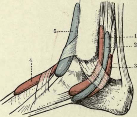
Fig. 44.-The Synovial Sheaths at the Ankle. Internal View. (After L. Testut's "Anatomie Humaine.")
11 Tibialis posticus. | 4. Extensor proprius hallucis |
2. Flexor longus digitorum. | 5. Tibialis amicus. |
3. Flexor longus hallucis. |
Continue to: