The Flexor Synovial Sheaths
Description
This section is from the book "Landmarks And Surface Markings Of The Human Body", by Louis Bathe Rawling. Also available from Amazon: Landmarks and Surface Markings of the Human Body.
The Flexor Synovial Sheaths
The flexor longus pollicis, the flexor sublimis and the flexor profundus digitorum all pass beneath the anterior annular ligament. Fig. XI.
In this situation the flexor sublimis consists of four tendons, of which the medius and annularis lie superficial to the tendons which pass to the index and little fingers. The profundus consists of two parts only, the tendon to the index-finger being alone differentiated off from the main mass. Beneath the ligament these tendons are surrounded by two synovial sheaths, one for the flexor longus pollicis and one for the remaining tendons plus the median nerve. The sheaths extend upwards about 1 inch above the upper limit of the ligament, and therefore the same distance above the lower transverse crease in front of the wrist. (Fig. XI, 6,6)
The flexor longus pollicis sheath is continued downwards to the insertion of the tendon into the distal phalanx of the thumb. The main sheath broadens out below the ligament and though generally continued onwards to the end of the little finger, (Fig. XI, 7,9) the major portion terminates at the level of the upper transverse crease of the palm. The flexor tendons to the fore, middle and ring fingers also possess more distally distinct synovial sheaths, (Fig. XI, 8.) which extend from the terminal phalanges of the fingers upwards to the necks of the metacarpal bones, a level corresponding roughly to the lower transverse crease of the palm.
A distance of 1/2 inch separates the main synovial sheath above from the more distal segments below.
On the outer side of the wrist the most marked feature is the "anatomical snuff-box," a space bounded on the radial side by the tendons of the extensor ossis metacarpi and primi internodii pollicis muscles, and on the ulnar side by the tendon of the extensor secundi internodii pollicis.
In the floor of the space the styloid process of the radius is felt, this prominence lying fully 1/2 inch below the level of the corresponding process of the ulna, and also on a slightly more anterior plane. Immediately below the radial styloid process the scaphoid bone lies, most prominent when the hand is well adducted. Below this, again, the trapezium and the bases of the first and second metacarpals are to be felt.
On the dorsum of the hand there is a well-marked elevation, most noticeable when the wrist is fully flexed, due to the projection of the bases of the second and third metacarpal bones, the styloid process of the latter bone being especially prominent.
Immediately above this elevation there is a depression where the tendons of the extensor carpi radialis longior and brevior are felt as they pass to their insertion into the bases of the second and third metacarpal bones.
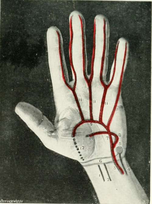
Fig. X. The Palm Of The Hand
1. The flexor carpi radialis.
2. The palmaris longus.
3. The ulnar artery.
4. The flexor carpi ulnaris.
5. The pisiform bone.
6. The transverse creases of the wrist
7. The superficial branch of the ulnar artery.
8. The deep branch of the ulnar artery.
9. The superficial palmar arch.
10. The deep palmar arch.
11. The digital branches of the superficial palmar arch.
12. The superficialis volae.
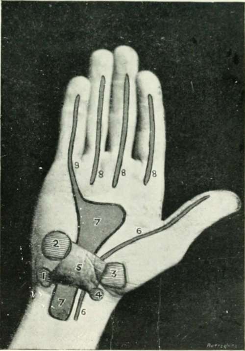
Fig. XI. The Palm Of The Hand
1. The pisiform.
2. The unciform
3. The trapezium.
4. The scaphoid tuberosity.
5. The anterior annular ligament.
6.The flexor longus pollicis sheath.
7. The main flexor synovial sheath.
8. The distal flexor synovial sheaths.
9. The continuation of the main sheath along the little finger.
Near the middle of the posterior aspect of the lower end of the radius a tubercle can generally be distinguished, (Fig. XII, 4.) the radial tubercle, separating the extensor secundi internodii pollicis on the inner side from the tendon of the extensor carpi radialis brevior, which lies more external.
The posterior annular ligament, about 3/4 inch broad, extends from the lower part of the outer border of the radius to the styloid process of the ulna and the carpal bones below the ulna. (Fig. XII, 1.)
The ligament has, therefore, a downward and inward direction, and beneath it pass the extensor tendons. These occupy distinct compartments, and possess synovial sheaths as under :
1. One compartment and synovial sheath for the extensor ossis metacarpi and extensor primi internodii pollicis. (Fig. XII, 2.)
2. One for the extensor carpi radialis longior and brevior. (Fig. XII, 3.)
3. One for the extensor secundi internodii pollicis. (Fig. XII, 5.)
4. One for the extensor communis digitorum and extensor indicis. (Fig. XII, 6.)
5. One for the extensor minimi digiti. Fig. XII,7. 6. One for the extensor carpi ulnaris. (Fig. XII, 8.)
The extent of the synovial sheaths is indicated in the diagram, where the radial artery is also depicted as it crosses the " anatomical snuff-box" towards the base of the first interosseous space, (Fig. XII, 9,10) at which level the vessel dips down between the two heads of the first dorsal interosseous muscle to complete the deep palmar arch.
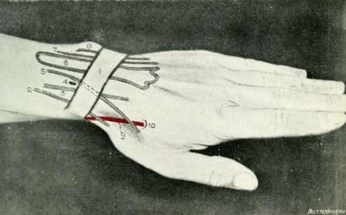
Fig. XII. The Back Of The Wrist
1. The posterior annular ligament. 4. The radial tubercle.
2, 3, 5-8. The compartments and synovial sheaths of the extensor tendons (see text).
9. The radial artery, crossing the " anatomical snuff-box."
10. The base of the first interosseous space.
Continue to: