The Common And Superficial Femoral Arteries
Description
This section is from the book "Landmarks And Surface Markings Of The Human Body", by Louis Bathe Rawling. Also available from Amazon: Landmarks and Surface Markings of the Human Body.
The Common And Superficial Femoral Arteries
With the thigh flexed, everted, and slightly abducted, these vessels correspond in direction to a line drawn from a point midway between the anterior superior iliac spine and the symphysis pubis to the adductor tubercle of the femur below.
The upper 1 1/2 inches of this line = the common femoral artery. (Fig XXIV, 6.)
The upper third = the common and superficial femoral arteries in Scarpa's triangle. (Fig. XXIV, 6,12)
The upper two-thirds = the complete common and superficial femoral arteries. (Fig. XXIV, 6, 12, 13)
The middle third = the superficial femoral artery in Hunter's canal. (Fig. XXIV, 13.)
The popliteal artery enters the upper angle of the popliteal space (from the inner side) by passing between the femur and the adductor magnus tendon. The vessel at first passes obliquely outwards and downwards to the mid-point of the space, and then changes direction by passing vertically downwards as far as the lower border of the popliteus muscle, at which level it bifurcates into anterior and posterior tibial arteries. The point of bifurcation corresponds to the level of the tubercle of the tibia.
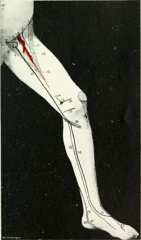
Fig. XXIV. The Side Of The Thigh And Leg
1. The anterior superior iliac spine.
2. The pubic spines.
3. The sartorius muscle.
4. The adductor longus muscle.
5. The anterior crural nerve.
6. The common femoral artery.
7. The common femoral vein.
8. The femoral ring.
9. The saphenous opening.
10.The internal or long saphenous vein.
11. The profunda femoris artery.
12. The superficial femoral in Scarpa's triangle.
13. The superficial femoral in Hunter's canal.
14. The adductor magnus tendon.
15. The adductor tubercle.
16. The lower epiphysial line of the femur.
17. The line of the knee-joint.
18. The gracilis, sartorius, and semitendinosus muscles.
19. The internal saphenous nerve.
20. The posterior tibial artery.
21. The internal plantar artery.
22. The external plantar artery.
23. The anterior tibial nerve.
Continue to:
- prev: The Vessels And Nerves Of The Lower Extremity
- Table of Contents
- next: The Anterior Tibial Artery