The Anterior Annular Ligament Of The Ankle
Description
This section is from the book "Landmarks And Surface Markings Of The Human Body", by Louis Bathe Rawling. Also available from Amazon: Landmarks and Surface Markings of the Human Body.
The Anterior Annular Ligament Of The Ankle
(Fig. XXVI, 3.) The upper portion of this ligament, about 1 inch broad, extends transversely across the ankle from tibia to fibula. It presents two compartments only, one for the tibialis anticus, (Fig. XXVI, 6.)and one for remaning extensor tendons. The former tendon alone possesses a synovial sheath.
The lower portion of the ligament is Y-shaped,(Fig. XXVI, 4,5) the single limb arising from the upper and outer aspect of the head of the os calcis in close connection with the origin of the extensor brevis digitorum muscle. The upper limb of the divided portion becomes attached to the internal malleolus, whilst the lower limb sweeps over to the scaphoid tuberosity and to the inner side of the foot. The extensor communis digitorum and the peroneus tertius pass under the single undivided limb, (Fig. XXVI, 8.) and possess in this situation a common synovial sheath; whilst the extensor longus hallucis and the tibialis anticus pass through separate compartments in each limb of the divided portion of the ligament, (Fig. XXVI, 6,7) and each tendon in so doing is surrounded by a synovial sheath, that enveloping the tibialis anticus tendon being continuous with the sheath already alluded to as enclosing the tendon under the transverse portion of the ligament.
The internal annular ligament is triangular in shape, (Fig. XXVII, 6.) the apex being attached to the internal malleolus, and the base to the lower margin of the os calcis. From the deep aspect of the ligament septa are given off which form separate compartments for the passage of the tendons of the tibialis posticus, (Fig. XXVII, 7,8,9) flexor longus digitorum, and flexor longus hallucis muscles, each tendon having its own synovial sheath. These three sheaths extend for about 1 inch above the upper limit of the annular ligament; and although the sheath enveloping the tibialis posticus reaches almost as far forwards as the scaphoid tuberosity, (Fig. XXIX, 3.) the other two sheaths usually terminate about 1/2 inch below the inferior margin of the ligament.
The flexor longus hallucis and flexor longus digitorum have, again, distinct synovial sheaths just before their insertion into the distal phalanges of the toes, these sheaths, however, being very variable, and rarely extending further backwards than the heads of the metatarsal bones.
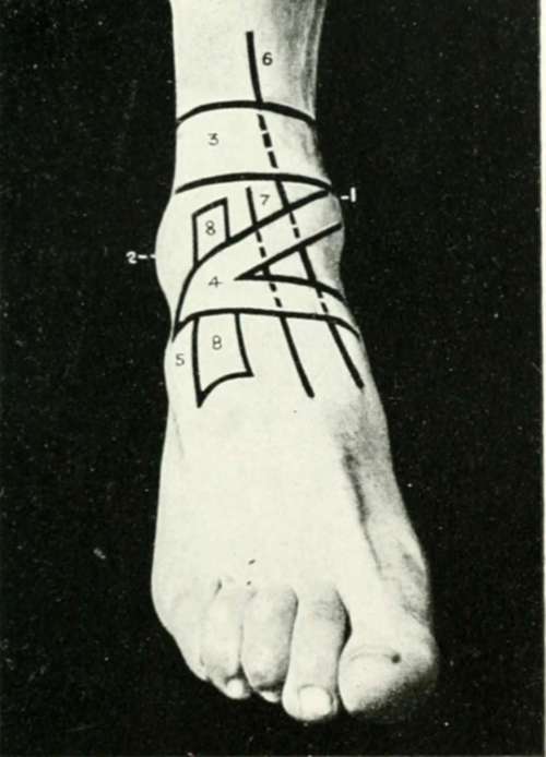
Fig. XXVI. The Region Of The Ankle And Foot
1. The internal malleolus.
2. The external malleolus.
3. The transverse band of the anterior annular ligament.
4. The Y-shaped hand of the anterior annular ligament.
5. The head of theos caicisand the extensor brevis digitortim.
6. The tibialis anticus synovial sheath.
7. The extensor longus hallucis synovial sheath.
8. The extensor longus digitorum and peroneus tertius synovial sheath.
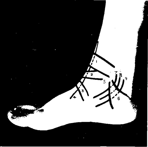
Fig. XXVII. The Region Of The Ankle And Foot
1. The transverse band.
2. 3. The upper and lower limbs of the Y-shaped part of the anterior annular ligament
4. The tibialis anticus synovial sheath.
5. The internal malleolus.
6. The internal annular ligament.
7. The tibialis posticus synovial sheath.
8. The flexus longus digitorum synovial sheath.
9. The flexor longus hallucis synovial sheath.
The external annular ligament is less definite in shape, and can only be described as a broad band passing from the external malleolus to the lower margin of the os calcis. (Fig. XXVIII, 4,5,6)
Beneath it two tendons pass, the peroneus longus and brevis. These two tendons possess a common synovial sheath, which extends upwards 2 to 3 inches above the tip of the external malleolus, and downwards as far as the "peroneal tubercle," where the sac divides into two, one part accompanying the peroneus brevis to near the base of the fifth metatarsal bone, the other extending forwards to the outer and under aspect of the cuboid bone. The peroneus longus is also usually enclosed in a synovial sheath in the last inch or so of its course, previous to its insertion into the outer aspect of the base of the first metatarsal bone.
Continue to: