Inferior Cervical Ganglion
Description
This section is from the book "Nerves Of The Human Body", by Charles R. Whittaker. Also available from Amazon: Hughes Nerves Of The Human Body.
Inferior Cervical Ganglion
A ganglion of considerable size nestling behind the first part of the subclavian artery, and between the transverse process of the seventh cervical vertebra and the neck of the first rib. It is incompletely separated from the first thoracic ganglion.
Branches
(a) Central. (1) Grey rami communicantes, to the anterior primary divisions of the seventh and eighth cervical nerves. (2) Ansa subclavia, to middle cervical ganglion.
(b) Peripheral. (1) Inferior cardiac, to the deep cardiac plexus.
(2) Vertebral, to vertebral plexus.
(3) Subclavian, to subclavian artery; it communicates with the phrenic nerve.
Functional Distribution
(a) Pupillo-dilator fibres (T. 1, 2, 3), run in the ascending branch of the superior cervical ganglion, pass to the Gasserian ganglion, and from thence along the ophthalmic division of the trigeminal, and the long ciliary nerves.
(b) Vaso-constrictor fibres.
(c) Secretory, to salivary glands (T. 2, 3),
(d) Accelerator fibres of heart (T. 2, 3, 4).
(e) Pilo-motor fibres of face and neck.
Thoracic Portion Of Sympathetic
The thoracic portion of the sympathetic rests upon the necks of the ribs and the intercostal vessels and nerves. It is clothed by the parietal layer of the pleura. There are usually ten or eleven ganglia
Branches
(a) Central. (1) White rami communicantes, from the anterior primary divisions of the lower eleven thoracic nerves. Those of the upper five are distributed to the head and neck, while the remainder are connected with the thoracic and abdominal viscera.
(2) Grey rami communicantes, pass to the anterior primary divisions of all the thoracic nerves.
(b) Peripheral. (1) Pulmonary, to the posterior pulmonary plexus from opposite the second, third, and fourth ganglia.
(2) Aortic, to the aorta from the first five ganglia.
(3) Great splanchnic, arises from the gangliated cord between the fifth or sixth and ninth ganglia. The nerve pierces the crus of the diaphgram to terminate in the semilunar (coeliac) ganglion.
(4) Lesser splanchnic, springs from the gangliated cord opposite the ninth and tenth ganglia, and enters the abdomen along with the great splanchnic, to end in the aortico-renal ganglion.
(5) Least splanchnic, emerges from the last thoracic ganglion. It passes through the diaphragm along with the other splanchnics, but enters the renal plexus.
Functional Distribution
(a) Vaso-constrictor fibres of limbs.
(b) Vaso-constrictor fibres of pulmonary vessels.
(c) Pilo-motor fibres of limbs.
Plate XI. Cutaneous Areas Of Upper Extremities
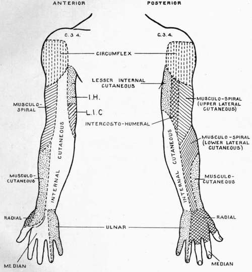
I.H. Intercosto-Humeral. L.I.C. Lesser Internal Cutaneous.
(d) Vaso-motor fibres of abdominal blood-vessels.
(e) Vaso-inhibitory fibres of stomach and intestines. (f) Constrictor fibres to the spleen.
(g) Motor fibres to musculature of rectum.
Continue to: