Chapter II. The Human Brain
Description
This section is from the book "Man: A History Of The Human Body", by Arthur Keith, Sir. Also available from Amazon: Man: A History Of The Human Body.
Chapter II. The Human Brain
We propose to visit the dissecting room again, this time with the definite purpose of seeing the human brainthat wonderful organ which has lifted mankind to so high an estate. We shall see a structure which, to all outward appearance, might have been that of a great manone of those who have written our plays, our novels, our philosophies, or who have conquered the world by force, by invention or by sweet persuasion. Here, on the threshold of the medical school, a confession must be made of our ignorance: no one, however skilled he may be, can tell, from merely surveying the brain, whether its owner was a clever man or a foolish one. The day may come when an examination of the brain will reveal its capacities, but we have not yet reached that position. Having made this confession we make our way to a small anatomical theatre in the school, and take a seat with the students on the benches which rise tier on tier, almost to the roof. Before us stands a white-coated anatomist; he is to give a demonstration on the brain, showing it to us in its natural position within the body. The scalp, in the subject of demonstration, is folded back, and the cap of the skull removed. Before the brain can be seen, a thick membrane (dura mater), which lines the interior of the skull or cranial cavity, has also to be turned aside, but this is easily done, for the brain is merely in contact with it. The chief part of the brain is thus exposedthe part known as the cerebrum. We note that it occupies that region of the skull which lies above the ear-holes, and that it extends forwards to occupy the forehead above the orbits and backwards to fill the projecting occiput above the root of the neck. The brain is wrapped in a semi-transparent membrane through which we see a rich supply of blood vessels on its folded or convoluted surface. Between the folds and convolutions there are depressions and fissures.
The cerebrum is divided into two hemispheres, right and left, a fact which we could not have guessed from our inner consciousness, for we seem to think as if with one organ. The anatomist presses the right and left halves slightly apart and shows a wide white bridge of nerve fibres (the corpus callosum) joining them. He further informs us of another mysterythe right hemisphere presides over the left half of the body while the left hemisphere is connected with the right side. He narrates a case, to illustrate the crossed relationship of brain and body, which he had seen some days previously. The case was that of a working-man, the subject of fits, in each of which he fell down with the fingers of the right hand moving convulsively. The surgeon marked out a small area on the left side of the patient's head, and operating there found a small tumour pressing on the underlying part of the brain. Its removal effected a cure. We see then why the surgeon operated on the left hemisphere when it was the right hand which was affected, but to realize how he was guided to the exact spot for operation we must look at the surface of the brain exposed before us. The surface is thrown into convolutions separated from each other by depressions. The longer and deeper depressions are known as fissures. We notice that the main onethe fissure of Sylviusbegins on that part of the brain which lies just behind the eye and beneath the temple and passes backwards and upwards to end some distance above the position of the tip of the ear (Fig. 1). The fissure of Sylvius separates the frontal and parietal lobes of the brain, situated above the fissure, from the temporal lobe, which is situated below it. Towards the posterior end of the brain the temporal and parietal lobes join with another important lobethe occipital. Crossing the surface of the hemisphere from above downwards and separating the frontal from the parietal lobe is a fissure of the utmost importance to us. It is the fissure of Rolando or central fissure (Fig. 1). On the convolution in front of the central fissurethe precentral convolutionis situated the area which regulates the movement of the arm. It was the knowledge of this fact which guided the surgeon. The central fissure is also seen on the brains of monkeys; in the manlike apes or anthropoids the fissure is very human-like. Some forty years ago it was discovered that when the cortex or surface layer of the convolution in front of the central fissure was stimulated electrically, the unconscious ape performed certain definite movements. If the upper part of the pre-central convolution was thus called into action certain movements of the opposite lower limb resulted; if the middle part, motions in the opposite upper limb ; if of the lower part, movements of the opposite side of the face, lips and tongue could be called forth. Further, it was discovered that the parts of the convolution behind the central fissure were connected with sensations arising in corresponding parts of the body. There are two inferences which we draw from these facts;
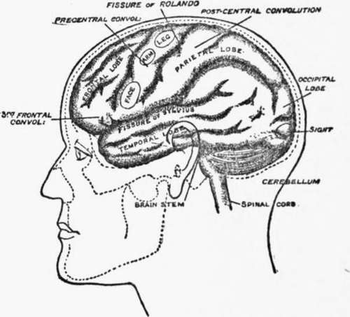
Fig. 1. The main divisions of the Brain, showing some of the functional centres.
(i) that the human brain must be formed on the same type or plan as that of the ape, for the medical man is able to apply, in diagnosing diseases of the brain, the knowledge he has obtained by experiments on the ape;
(ii) that each part of the brain has its own peculiar function. Only the convolutions immediately in front of the central fissure give rise to movements ; when other parts are stimulated there is no muscular response. Thus it is, that at the present time there are large parts of the brain whose function is unknownparts which we believe serve for memory, judgment and imagination.
We must, however, keep our eyes on the demonstrator and watch him as he proceeds to show us how the cerebral hemispheres are connected with the body. When they are raised from the floor or base of the skull we see a great stemthe brain stemissuing from them ; in size it is about the thickness of a baby's wrist. When this is cut through the cerebrum can be lifted away, and we note the remarkable fact that only one pair of nerves end directly in the hemispheres, namely, the olfactory or nerves of smell. The optic nerves from the eye are now visible ; they encircle and seem to end on the back of the brain stem. Impressions arising in the eyethe sensations caused by lightpass first to the brain-stem, but from their termination other nerve paths are provided which can be traced into the cerebral hemisphere towards the posterior or occipital lobes. The occipital cortex in which the paths end are known to subserve the functions of sight; blindness is produced when they are destroyed by disease. When the cerebral hemispheres are removed from the head it is seen that their posterior parts did not rest on the base of the skull as the frontal and temporal lobes did, but on a fibrous partition. When the anatomist removes the partition we see beneath it a minor compartment which contains the cerebellum and the continuation of the stem of the brain.
Continue to:
Tags
human body, brain, human sexuality, muscles, anthropoids, human birth