The Structure Of The Thorax
Description
This section is from the book "The Human Body: An Elementary Text-Book Of Anatomy, Physiology, And Hygiene", by H. Newell Martin. Also available from Amazon: The Human Body.
The Structure Of The Thorax
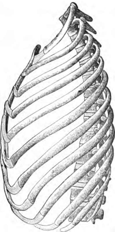
The thorax is a conical cavity with a rigid supporting skeleton (Pig. 68) formed by the dorsal vertebra behind, the breast bone in front, and the ribs and rib cartilages on the sides. Between and over these lie muscles, and the whole is covered air-tight by the skin outside and the pleura (p. 266) inside. Above, it is closed by the muscles and other organs of the neck; and below by a movable bottom, the diaphragm. The air-tight chamber thus bounded can be enlarged in all three diameters, but especially in the vertical, and in that running from the spinal column to the breastbone (dorso-ventral diameter).
The Vertical Enlargement Of The Thorax
This is brought about by the contraction of the diaphragm, which (as may be seen in Fig. 69), is a thin sheet of muscle, with a fibrous membrane in its centre serving as a tendon. In rest the diaphragm is dome-shaped, its concavity being turned towards the abdomen. From the tendon on the crown of the dome, striped muscular fibres radiate, downwards and outwards, in all directions, and are fixed by their inferior ends to the lower ribs, the breast-bone, and the vertebral column. In inspiration (drawing a breath) the muscular fibres, shortening, flatten the dome and so enlarge the thoracic cavity at the expense of the abdominal. The contraction of the diaphragm thus greatly increases the size of the thorax chamber by adding to its lowest and widest part.
What when we breathe out?
What is inspiration? Expiration? How often does each occur in a minute? Compare the rate of respiration and of the heart-beat?
What is the form of the thorax? What forms its skeleton? What lie between and over its bones and cartilage? How is it closed airtight?
How is the chest closed above? How below? In what diameters can the chest cavity be enlarged? Describe the diaphragm.
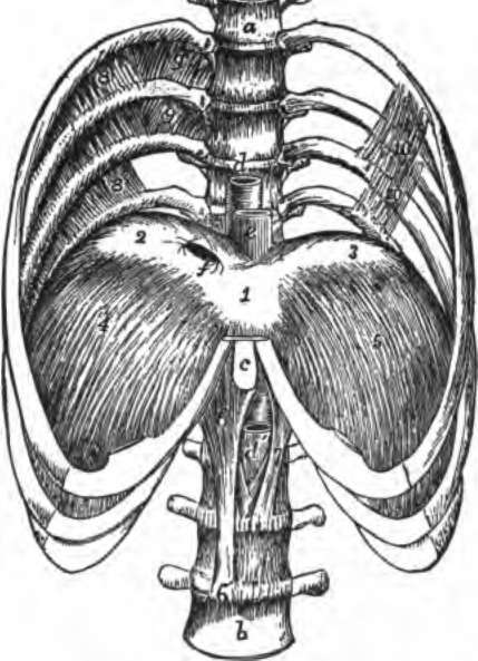
Fig. 69. The lower half of the thorax, with four lumbar vertebra, showing the diaphragm from above. 1,2, 3, central tendon; 4, 5, muscular part.
What happens to it during inspiration? What results?
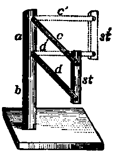
Fig. 70. Diagram illustrating the dorso ventral increase in the diameter of the thorax when the ribs are raised.
The Dorso Ventral Enlargement Of The Thorax
The ribs on the whole slope downwards (Fig. 15) from the vertebral column to the breast-bone, the slope being most marked in the lower ones. During inspiration the breast-bone and the sternal ends of the ribs attached to it are raised by muscles which pull on them, and so the distance between the sternum and the vertebral column is increased. That this must be so will readily be seen by examining the diagram Fig 70, where ab represents the vertebral column, c and d two ribs, and st the sternum. The continuous lines represent the natural position of the ribs at rest in expiration, and the dotted lines the position in inspiration. It is clear that when their lower ends are raised so as to make the bars lie in a more horizontal plane, the sternum is pushed away from the spine, and so the extent of the chest cavity between back-bone and breast-bone is increased.
Continue to: