The Spinal Cord
Description
This section is from the book "The Human Body: An Elementary Text-Book Of Anatomy, Physiology, And Hygiene", by H. Newell Martin. Also available from Amazon: The Human Body.
The Spinal Cord
The Spinal Cord (Fig. 80) is nearly cylindrical in form, being, however, a little wider from side to side than dorso-ventrally, and tapering off at its posterior end. Its average diameter is about 3/4 inch and its length 17 inches. It weighs 1 1/2 ounces. There is no marked limit between the spinal cord and the brain, the one passing gradually into the other. In its course the cord presents two expansions, an upper, 10 (Fig. 80), the cervical enlargement, reaching from the third cervical to the first dorsal vertebræ, and a lower or lumbar enlargement, 9, opposite the last dorsal vertebræ.
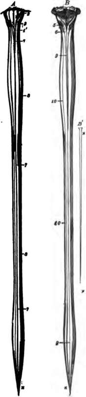
Fig. 80. The spinal cord and medulla oblongata. A, from the ventral, and B, from the dorsal aspect; C to H, cross-sections at different levels.
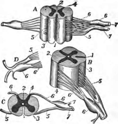
Fig. 81. The spinal cord and nerve-roots. A, a small portion of the cord seen from the ventral side; B, the same seen laterally; C, a cross-section of the cord; D, the two roots of a spinal nerve; 1, anterior (ventral) fissure; 2, posterior (dorsal) fissure; 3, surface groove along the line of attachment of the anterior nerve-roots; 4, line of origin of the posterior roots; 5, anterior root filaments of a spinal nerve; 6, posterior root filaments; 6', ganglion of the posterior root; 7, 7', the first two divisions of the nerve-trunk after its formation by the union of the two roots.
What is the arachnoid? What is the cerebro-spinal liquid? What is the general form of the spinal cord? Its average diameter? Its length? Its weight? How does it connect with the brain?
Running along the middle line on both the ventral and the dorsal aspects of the cord are fissures which (0, Fig. 81) nearly divide it into right and left halves.
A transverse section, C, shows that the substance of the cord is not alike throughout, but that its white superficial layers envelop a central gray substance arranged somewhat in the form of a capital H. Each half of the gray matter is crescent-shaped, and the crescents are turned back to back and united across the middle line by the gray commissure.
The Spinal Nerves
Thirty-one pairs of spinal nerves join the spinal cord in the neural canal of the vertebral column, entering through the intervertebral foramina (p. 44). Each divides in the foramen into a dorsal and ventral portion, often named the posterior and anterior roots of the nerve (6 and 5, Fig. 81), and these are attached to the sides of the cord. On each dorsal root is a spinal ganglion (6', Fig. 81), placed where it joins the anterior or ventral root to form the common nerve-trunk. Immediately after the mixture of fibres from both roots, the trunk begins to divide into branches for the supply of some region of the body.
What expansions are seen in it? Where is each placed?
How are the right and left halves of the cord separated?
Is the spinal cord alike all through? What are the colors of its outer and of its inner portions? What is the form of the gray matter of the cord as seen on cross-sections? How are the crescents united?
How many spinal nerves are there? What do they join? How do they get into" the neural canal? Where do they divide? What are the divisions named? To what are they attached? What is found on each posterior root?
Continue to: