The Lachrymal Apparatus
Description
This section is from the book "The Human Body: An Elementary Text-Book Of Anatomy, Physiology, And Hygiene", by H. Newell Martin. Also available from Amazon: The Human Body.
The Lachrymal Apparatus
The Lachrymal Apparatus consists of the tear-gland in each orbit, of ducts which carry its secretion to the upper eyelid, and of canals, by which this, unless when excessive, is carried off from the front of the eye without running down over the face. The lachrymal or tear gland, about the size of an almond, lies in the upper and outer corner of the orbit. It is a compound racemose gland, from which twelve or fourteen ducts run and open at the outer corner of the upper eyelid on its inner surface. The secretion there poured out is spread evenly over the exposed part of the eye by the movements of winking, and keeps it moist; finally it is drained off by two lachrymal canals, one of which opens by a small pore on an elevation, or papilla, near the inner end of the margin of each eyelid. The aperture of the lower canal can be readily seen by examining its papilla in front of a looking-glass. The canals run inwards and open into the lachrymal sac, which lies just outside the nose, in a hollow where the lachrymal and superior maxillary bones (L and Mx, Fig. 16) meet. From this sac the nasal duct proceeds to open into the nose-chamber below the inferior turbinate bone (q, Fig. 41, p. 151).
Tears are constantly being secreted, but ordinarily in such quantity as, to be drained off into the nose, from which they flow into the pharynx and are swallowed.
When the lachrymal duct is stopped up, however; their continual presence makes itself unpleasantly felt, and may need the aid of a surgeon to clear the passage. In weeping the secretion is increased, and then not only more of it enters the nose, but some flows down the cheeks. The frequent swallowing movements of a crying child, sometimes spoken of as "gulping down his passion,"are due to the need of swallowing the extra tears which reach the pharynx.
Of what parts does the lachrymal apparatus consist ? Describe the lachrymal gland. Where do its ducts open ? How is the front of the eyeball kept moist ? Describe the arrangement by which the tears are usually carried off.
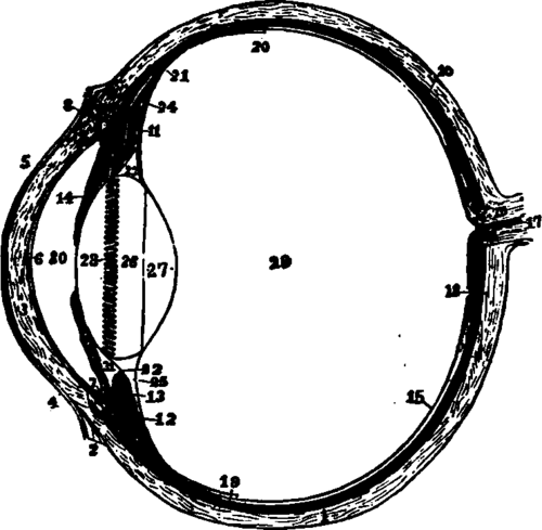
Fig. 88. The left eyeball in horizontal section from before back. 1, sclerotic; 2, junction of sclerotic and cornea; 8, cornea: 4, 5, conjunctiva; 6, posterior elastic layer of cornea; 7, ciliary muscle; 10, choroid; 11. 13, ciliary processes; 14, iris; 15, retina; 16, optic nerve; 17, artery entering retina in optic nerve; 18, fovea centralis; 19, region where sensory part of retina ends: 22, suspensory ligament; 23 is placed in the canal of Petit, and the line from 26 points to it; 24, the anterior part of the hyaloid membrane; 26, 27, 28, are placed on the lens; 28 points to the fine of attachment around it of the suspensory ligament: 29, vitreous humor; 30, anterior chamber of aqueous humor; 31, posterior chamber of aqueous humor.
Why do tears run down the face during a fit of weeping?
Why does a crying child make frequent swallowing movements?
Continue to: