The Histology Of Bone
Description
This section is from the book "The Human Body: An Elementary Text-Book Of Anatomy, Physiology, And Hygiene", by H. Newell Martin. Also available from Amazon: The Human Body.
The Histology Of Bone
The microscope shows that compact bone is only so to the naked eye ; even a hand lens shows minute holes in it; it but differs from spongy bone in the fact that its cavities are much smaller, and the hard bony plates between them thicker. It a thin transverse section of the shaft of long bone (Fig. 23) be examined with a microscope magnifying about twenty diameters, even its densest part will be seen to show numerous openings which become gradually larger near the medullary cavity and pass insensibly into the spaces of the spongy bone around it. These openings are the cross sections of tubes known as the Haversian canals, which run all through the bone, the majority of them in the direction of its long axis, though numerous cross branches unite them. The outermost Haversian canals open on the surface of the bone beneath the periosteum; from there blood-vessels pass in, and, traversing the whole bone in these channels, convey materials for its growth and nourishment.
What is a long bone? Give examples. A tabular bone? Examples. A short bone? Examples. An irregular bone? Examples.
With what is most of the surface of bones covered? Where is the periosteum absent? What bones contain yellow marrow? What do the others contain?
Does compact bone contain any cavities? How may these be seen ? How does it differ from spongy bone ? What is seen when a thin slice of bone is magnified twenty times ? Where do the apertures in it become larger?
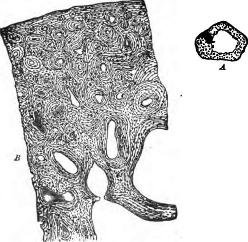
Fig. 23. A, a transverse section of the ulna, natural size; showing the medullary cavity. B, the more deeply shaded part of A magnified twenty diameters.
What are the Haversian canals? Where do the outer ones open? What enters them? Where do the blood-vessels of a bone run? What do they carry?
Around each Haversian canal is a series of plates or lamell, each canal and its lamell forming an Haversian system ; the entire bone is made up of a number of such systems, with the addition of a few lamell lying in the corners between them, and some which run around the whole bone on its outer surface. In the spongy bone the Haversian canals are very large, containing red marrow as well as blood-vessels, and the lamell around each are few in number.
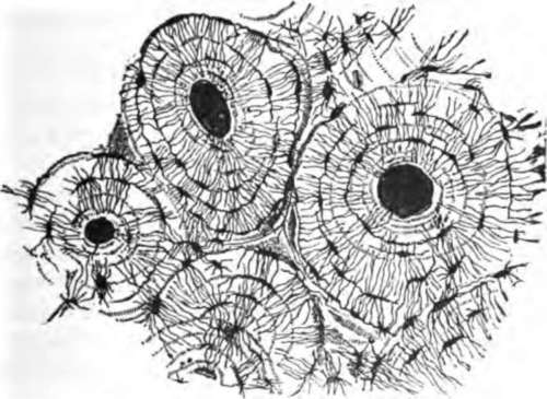
Fig. 24. A email piece of bone, ground very thin and highly magnified.
If a bit of bone be still more magnified (Fig. 24) we find that very small cavities lie between the lamell; they are called lacun; from each lacuna radiate many extremely fine tubes, the canaliculi, so that each lacuna with its canaliculi looks something like a small animal with a great many legs. The innermost canaliculi open into the Haversian canal of the system to which they belong, and those of various lacun communicate with one another, so that a set of passages is provided through which liquid which transudes from the blood-vessel in the Haversian canal can ooze all through the bone.
What lies around each Haversian canal? What is an Haversian system? Of what does a bone consist in addition to Haversian systems? Arc the Haversian canals comparatively large or small in spongy bone? What do the spaces of spongy bone contain in addition to blood-vessels?
What spaces he between the lamell of an Haversian system?
In a living bone a nucleated cell lies in each lacuna. These cells, the bone corpuscles, are the remnants of those which made the bone, all whose hard parts are but intercellular substance: a sort of skeleton is made by each cell around itself, and this adheres to the skeletons of the rest, and thus the whole bone is built.
Continue to:
- prev: Varieties Of Structure Found In Different Bones
- Table of Contents
- next: Chemical Composition Of Human Bone