The Course Of The Main Arteries Of The Body
Description
This section is from the book "The Human Body: An Elementary Text-Book Of Anatomy, Physiology, And Hygiene", by H. Newell Martin. Also available from Amazon: The Human Body.
The Course Of The Main Arteries Of The Body
The aorta after leaving the left ventricle makes an arch (αA.) with its convexity towards the head. From the heart end of this arch arise the coronary arteries, which carry blood into the walls of the heart. From the convexity of the arch spring the three large trunks: the innominate artery ; the left common carotid artery (cs); and the left subclavian artery (Ssi). The innominate soon divides into the right subclavian (sd), and the right common carotid (cd) Each common carotid runs up the neck on its own side, and divides into branches for the neck, face, scalp, and brain. Each subclavian continues across the arm-pit as the axillary artery (ax), and then runs down to near the elbow as the brachial artery (b). Just above the elbow it divides into the radial and ulnar arteries (r, u,) which supply the fore-arm, and end in small branches for the hand.
What is found between each auricle and ventricle? What are they called? Describe the mitral valve. Where is it placed? What are the chordæ tendineæ? The papillary muscles? How far will the cords allow the valve flaps to rise? How does the tricuspid valve differ from the mitral?
How many semilunar valves are there? Where are they placed? Describe them. What is their use?
Beyond its arch the aorta runs back close to the spinal column as the thoracic aorta (At), which gives off branches to the walls of the chest and some organs in that cavity. The vessel then passes through the diaphragm, and continues as the abdominal aorta (Aαb) to the lower part of the abdomen. The main branches of the abdominal aorta are: (1) the cliac axis, which divides into branches for the stomach, liver, and spleen; (2) the upper and lower mesenteric arteries, which supply the intestines with blood; (3) the renal arteries (k), which carry blood to the kidneys.
What is meant by the aortic arch? What are its first branches? What branches are given off from the upper side of the aortic arch? Into what vessels does the innominate artery divide? To what parts do the common carotid arteries carry blood? In what does the subclavian artery terminate? What vessels supply the fore-arm with blood? How do they end?
What is the thoracic aorta?
The abdominal aorta? Name the chief branches of the abdominal aorta. What organs are supplied by the cliac axis? The mesenteric arteries? The renal arteries?
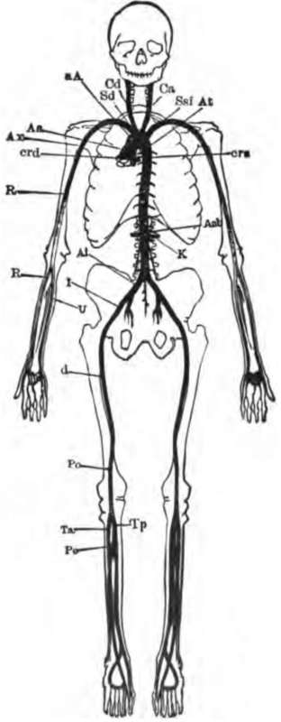
Fig. 60. The main arteries of the body. Crd and Crs, right and left coronary arteries of the heart, cut short near their origin; Aα, and αA, aortic arch; At, thoracic aorta; Aαb, abdominal aorta; K, renal artery; Sd, right, and Ssi. left subclavian: Cd, right, and Cs. left carotid; Ax. axillary artery; B, brachial artery; U, ulnar artery; R, radial artery; Ai, common iliac artery; J. external iliac artery; C, femoral artery; Po, popliteal artery; Ta, anterior, and Tp, posterior tibial artery; Pe, peroneal artery.
The two trunks into which the posterior end of the abdominal aorta divides (Ai) are named the common iliac arteries; each gives off some branches in the pelvis, and then continues along the thigh as the femoral artery (C); this runs to the knee-joint, behind which it is called the popliteal artery (Po). The popliteal artery divides into the peroneal (Pe) and tibial (Ta, Tp) arteries, which supply the lower leg and the foot.
The Properties Of The Arteries
Two fundamental facts must be borne in mind in connection with the arteries: First, that they are highly elastic and extensible; a large artery is, in this respect, much like a piece of rubber tubing of the same size. Second, the arteries have rings of muscular tissue in their walls, and when the muscle contracts the bore of the artery (and consequently the amount of blood which flows through it) is diminished. When the muscle relaxes, the bore of the artery is increased, and more blood passes along it to the capillaries in which it ends.
Continue to: