The Cavities Of The Heart
Description
This section is from the book "The Human Body: An Elementary Text-Book Of Anatomy, Physiology, And Hygiene", by H. Newell Martin. Also available from Amazon: The Human Body.
The Cavities Of The Heart
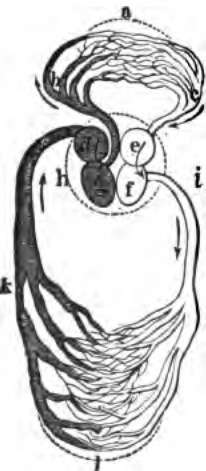
On opening the heart (see diagram, Fig. 57, p. 222) it is found to be subdivided by a longitudinal partition or septum into completely separated right and left halves, the partition running from about the middle of the base to a point a little on the right of the apex. Each of the chambers on the sides of the septum is again incompletely divided transversely into a thinner basal portion into which veins open, known as the auricle, and a thicker apical portion from which arteries arise and called the ventricle. The heart cavity thus consists of a right auricle and ventricle and a left auricle and ventricle, each auricle communicating by an auriculo-ventricular orifice with the ventricle on its own side; there is no direct communication whatever through the septum between the opposite sides of the heart. To get from one side to the other the blood must leave the heart and pass through a set of capillaries, as may readily be seen by tracing the course of the vessels in Fig. 57.
Why should a person with rheumatic fever never be exposed to cold except under skilled advice? What can be heard with a stethoscope in the early stages of pericarditis? What may occur in its later stages?
What is seen on opening the heart? In what direction does the septum run? What is an auricle? A ventricle? Enumerate the chambers of the heart cavity. With what does each auricle communicate? Is there a direct connection between the right and left sides of the heart? What must the blood do to get from one side of the heart to the other?
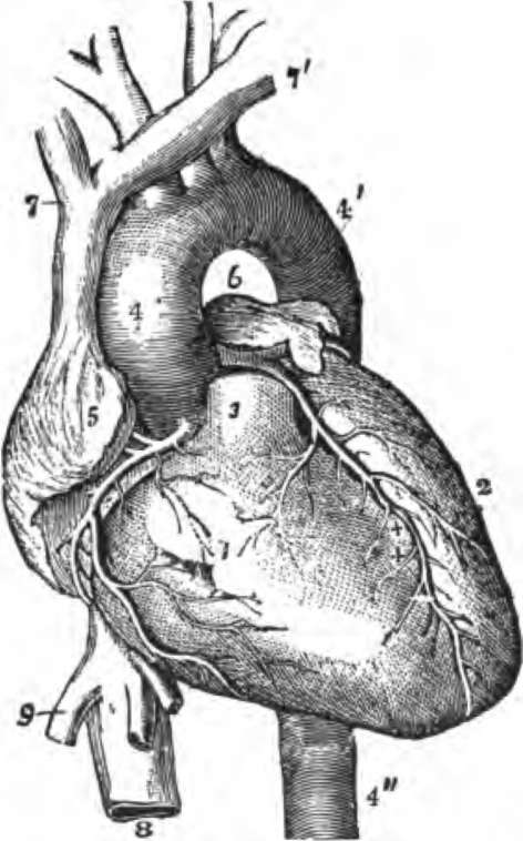
Fig. 58. View of the Heart and Great ve88els from before.
The pulmonary artery has been cat short close to its origin. 1, right ventricle; 2, left ventricle: 3, root of the pulmonary artery; 4, 4', arch of the aorta; 4", the descending thoracic aorta; 5. part of the right auricle; 6, part of the left auricle ; 7, 7', innominate veins joining to form the vena cava superior; 8, inferior vena cava, 9, one of the large hepatic veins; X, placed in the right auriculo-ventricular groove, points to the right or posterior coronary artery; X. X, placed In the anterior interventricular groove, indicate the left or anterior coronary artery.
Continue to:
- prev: The Position Of The Heart
- Table of Contents
- next: The Vessels Connected With The Different Chambers Of The Heart