87. The Arterial Valves
Description
This section is from the book "Animal Physiology: The Structure And Functions Of The Human Body", by John Cleland. Also available from Amazon: Animal Physiology, the Structure and Functions of the Human Body.
87. The Arterial Valves
The Arterial Valves guarding the entrances into the pulmonary artery and aorta are named semilunar, because they consist each of three delicate pouches, with semilunar attachments to the wall of the artery. The pouches are placed in a circle, with their mouths turned away from the heart, and are pushed fiat against the arterial walls when the blood is rushing out of the ventricles; but as soon as the ventricular contraction ceases, and the elasticity of the arteries tends to make the blood recoil, they are filled with the blood in the arteries, the sides of each are pressed against the adjacent sides of the two others, and all three reach in to the centre of the orifice, so as effectually to block it up. The action of these valves can be studied in a sheep's heart, by pouring water into the cut arteries, when it will be seen that not a drop passes back into the ventricles.
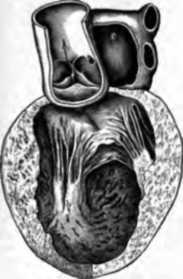
Fig. 66. Lett Side of the Heart.
The pulmonary artery has been removed. An arrow is passed through the aortic orifice between the semilunar poaches of its valve; and the lower end of the arrow rests on the anterior cusp of the mitral valve. a, b, Anterior and posterior musculi papillares, with chords tendineteæ passing up from each to both cusps; c, auricular cavity.
The auriculo-ventricular valves of the right and left sides of the heart are named respectively the tricuspid and the bicuspid or mitral, the one consisting of three pointed membranous flaps or cusps, and the other of two. The cusps are attached at the base to the margins of the auriculo-ventricular orifices, and are kept in the interior of the ventricles by a number of threads, chorda tendineae, attached to their edges and backs, and fastened at the other end to muscular prominences, the musculi papillares. The arrangements are on the same principle in both valves, but may bo studied best on the mitral, which is the more perfect of the two. The chordæe tendineæ of each cusp are divided into two sets; those from each half joining with those of the adjacent half of the other cusp to be inserted into one muscuius papillaris. Thus the contraction of the musculi papillares not only prevents the cusps from being flung into the auricle, but keeps their edges in apposition. These muscles contract at the same time as the ventricular wall with which their fibres are continuous, and the contraction of the ventricle pushes the blood against the backs of the valves, so as to bring them quite together, block up completely the passage into the auricle, and leave the blood no other aperture of exit save into the artery.
88. If we apply our ear to any one's chest, we find that the heart makes some noise in its action, that there is a perpetual " pit-pat, pit-pat," or recurrence of two successive sounds; first a slightly prolonged sound, then, a moment afterwards, another short and clear, and after that a longer interval before the first sound is repeated. The first sound will be found to occur at the same moment as the beat against the chest, and nearly at the same moment as the pulse at the wrist; it is synchronous also with the systole or contraction of the ventricles, and is caused by the vibration of the auriculo-ventricular valves when suddenly closed by the pressure of the blood on them. It continues audible in an animal when the chest has been laid open, so does the second. The second sound is caused by the closure of the arterial valves, as has been experimentally proved by introducing a pair of needles, one into the pulmonary artery, and the other into the aorta, so as to prevent the valves shutting, and observing that at once the sound ceases.
The beating of the heart against the chest is called the impulse, and is caused by the apex, which is at all times in contact with the wall of the chest, being pressed against it in the ventricular contraction by jerking upwards, forwards, and to the left. This movement is probably caused by the arch of the aorta being thrown into a rigid and more expanded curve when filled with blood, while it is fixed in its position behind; but it has been suggested that it results from the disposition of the muscular fibres of the ventricles.
Continue to: