193. The Internal Ear
Description
This section is from the book "Animal Physiology: The Structure And Functions Of The Human Body", by John Cleland. Also available from Amazon: Animal Physiology, the Structure and Functions of the Human Body.
193. The Internal Ear
The Internal Ear is imbedded in osseous tissue; and the cavity which it occupies, the osseous labyrinth, can be well studied in a macerated temporal bone. The petrous portion of the temporal bone is a long three-sided pyramid of hard consistence, with its base turned towards the tympanum; and in its posterior wall, which looks into the cranium, there is a large foramen, the meatus auditorius internus, directed outwards. When this meatus is looked into, it is seen to terminate very soon in a perforated plate, which occupies its lower part, and leaves an opening into a canal above it. This canal, termed aquaeductus Fallopii, gives passage to the portio dura of the seventh nerve, otherwise called the facial, and has nothing to do with the organ of hearing; but the perforated or cribriform plate transmits the portio mollis or auditory nerve to the internal ear; the perforations in its fore and hinder moiety leading respectively into the anterior and posterior divisions of the labyrinth. The posterior part of the osseous labyrinth is called the osseous vestibule, and contains the membranous vestibule floating within it; the anterior part is called the cochlea, and in it the osseous and membranous parts are more intimately connected one with the other. However, in the dry bone, the parts seen are the cavity of the vestibule, with three semicircular canals coming off from it behind; and, in front of the vestibule, the cochlea, a spiral tube coiled on itself like a snail's shell.
The cavity of the vestibule is the part into which the fenestra ovalis opens. The three semicircular canals spring from its back part; they are about a twentieth of an inch in width, and each makes a circuit about a quarter of an inch in diameter. One of them, the external, is placed horizontally; the other two, called the posterior and the superior, are somewhat larger, and lie in planes at right angles one to the other; the posterior being close to the hinder surface of the bone, and the superior springing from a part which is common to it and the posterior canal, and arching outwards, touching the upper surface of the bone. Each semicircular canal has a dilatation or ampulla near one extremity.
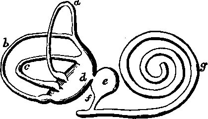
Fig. 129. Membranous Labrynth, diagrammatic view, a, b, c, Superior, posterior, and external semicircular canals; d, utricle; e, saccule; f, canalis reuniens; g, canalis membranacea cochleae.
The membranous vestibule, lodged within the osseous, is connected with it merely by vessels and nerves. It presents two cavities separated by a thin partition: the anterior is named the saccule, and connected, as we shall see, with the cochlea; the posterior, which is larger, is called the utricle, and gives off membranous semicircular canals,which lie loose in the osseous canals of the same name, and likewise exhibit each an ampulla. The fluid in which all these membranous structures float is called the perilymph, and that which they contain is called the endolymph. The nerves are supplied only to limited portions of the membranous vestibule, one branch passing to the saccule, another to the utricle, and one to a little crescentic elevation projecting into the interior of each ampulla. The interior of the membranous vestibule is lined with epithelium; and exceedingly slender straight hairs project through the epithelium at the parts supplied with nerves, and are probably in continuity with the nerve fibres. These hairs may be supposed to vibrate with sonorous vibrations; and to make such vibrations stronger, there is a collection of minute crystals of carbonate of lime, the otoliths or otoconia, in the neighbourhood of the termination of each branch of nerve.
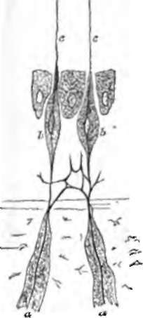
Fig. 130. Nerve-terminations in an Ampulla, diagrammatic view, a, a, Medullated nerve fibres; b,b, fusiform cells; c, c, auditory hairs. Rudinger.
Continue to: