145. The Whole Of The Brain Above The Level Of The Tentorium
Description
This section is from the book "Animal Physiology: The Structure And Functions Of The Human Body", by John Cleland. Also available from Amazon: Animal Physiology, the Structure and Functions of the Human Body.
145. The Whole Of The Brain Above The Level Of The Tentorium
The Whole Of The Brain Above The Level Of The Tentorium is included under the name of cerebrum, and is connected with the parts below by a neck or isthmus, in thickness about the size of a florin, which traverses a space left between the free edge of the tentorium and the body of the sphenoid bone. This isthmus, as seen from below, consists of two thick pillars, the crura cerebri, emerging above the pons Varolii, diverging as they ascend, and almost immediately concealed by the two cerebral hemispheres.
The cerebral hemispheres in man form by far the most bulky part of the brain. They are covered with a thick coating of grey matter of stratified structure, exhibiting an arrangement of corpuscles and fibres connecting one part of it with another, and the whole with the parts of the brain from which the hemispheres arise, as illustrated in fig. 101. This grey matter is thrown into a number of convolutions arranged on a definite plan, though varying in their finer details in different individuals, and even on the two sides of the brain. They are absent in the lower orders of mammals, become more abundant the higher we ascend in the series, and reach their greatest complexity in man. The only plausible explanation of the use of these convolutions is this, that grey matter requiring a rich vascular supply, and only minute vessels being admissible within it, increase of surface is a necessary condition of increase of bulk, so as to bring its parts sufficiently near the pia mater, in which the arteries for its nourishment divide. Nor is this supposition contradicted by the presence of convolutions in the brains of small animals; for in them the length of capillary travelled by the blood in each circulation is likewise small, and, therefore, the distance to which the blood can penetrate from the pia mater may be supposed to be in proportion to the size of the animal.
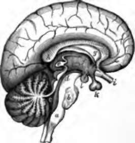
Fig. 100. Mesial Section of Brain. a, Medulla oblongata; b, pons Varolii ; c, fourth ventricle, and, behind it, valve of Vieussens; d, iter, and, behind it, corpora quad-rigemina ; e, pineal body; f, optic thalamus looking into the third ventricle, and, in front of it, an open passage from the third to the lateral ventricle, called foramen of Monro; g, left layer of septum lucidum bounding the fifth ventricle, and, beneath it, the fornix; h, posterior extremity of corpus callosum above the transverse fissure; i, optic nerve; k, pituitary body; 1, one of the corpora albicantia.
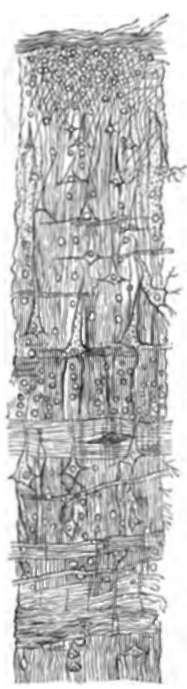
Fig. 101. Gray Matter of the Convolutions : ideal vertical section.
The hemispheres are in contact with very nearly the whole extent of the cranial wall above the tentorium. They are separated one from the other above, behind, and in front by a deep longitudinal fissure, into which dips a process of dura mater, called the falx cerebri, attached behind along the middle line of the tentorium, and in front to the mesial part of the ethmoid bone. At the bottom of the longitudinal fissure they are united by a thick transverse commissure or bond of junction, the corpus callosum, which presents a thick posterior border in front of the tentorial attachment of the falx, and at its fore part curves downwards behind the ethmoidal attachment of that structure, to become continuous with a thin lamina which completes the floor of the cavity of the brain in the middle line.
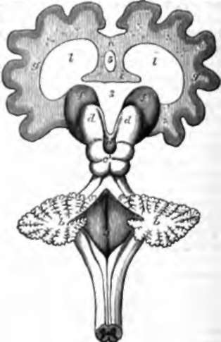
Fig. 102. Diagram of Brain, a. Spinal cord ; b, b, cerebellum divided, and, above it, the valve of Vieusaens partially divided; e, corpora quadrigemina; d, d, optic thalami; e, pineal body ; f, f, corpora striata; g, g, cerebral hemispheres in section; h, corpus callosum ; i, fornix; l, l, lateral ventricles ; 3, third ventricle; 4, fourth ventricle; 5, fifth ventricle, bounded on each side by septum lucidum.
The parts of the hemispheres, which project forwards, resting on the anterior fossa of the skull, over the orbits, are called anterior lobes; the parts above the cerebellum are the posterior lobes, and the parts turning down over the crura cerebri, and resting in the middle fossæ, on the sphenoid and temporal bones, are distinguished as the middle lobes.
146. If a human brain, or still better, the brain of any of the domestic quadrupeds, be examined, and the cerebellum be turned aside from the hemispheres, the isthmus will be brought into complete view, and there will be displayed above it an elevation divided by a crucial depression into four parts, and called on that account corpora quadrigemina. Two tracts will also be seen, about half an inch separate, adherent to the crura cerebri, and proceeding to the corpora quadrigemina from the cerebellum; these are the superior crura cerebelli already alluded to; and, between these, a thin lamina, called valve of Vieussens, limited by the cerebellum behind and the corpora quadrigemina in front, forms the roof of the fore part of the fourth ventricle, as that hollow is continued forwards into a narrow canal or iter which passes beneath the corpora quadrigemina, and opens in front of them.
By reflecting the hemispheres well forwards off the crura, a pair of large elevations, the optic thalami, will be exposed in front of the corpora quadrigemina; and, by dividing the corpus callosum and other structures, so as to permit more complete reflection of the hemispheres, still another pair of elevations, the corpora striata, will be seen in front of the optic thalami, and external to them. A soft body, about the size of a pea, the pineal body, will be likewise noticed attached by a slender connection in front of the corpora quadrigemina, and overhanging them, imbedded in pia mater. It may be mentioned of this structure that it is remarkable in being present in all the divisions of the vertebrata, although in the lower forms represented by little else than vascular tissue, and in man consisting of degenerated brain structure.
Continue to:
- prev: 144. Structure Of The Encephalon
- Table of Contents
- next: The Whole Of The Brain Above The Level Of The Tentorium. Continued