A Dissection To Expose The Peroneal Artery And Its Branches
Description
This section is from the book "A Manual Of Dissections Of The Human Body", by R. E. Carrington. Also available from Amazon: A manual of dissections of the human body.
A Dissection To Expose The Peroneal Artery And Its Branches
Position
The body supine, the leg rotated outwards and lying upon its inner aspect.
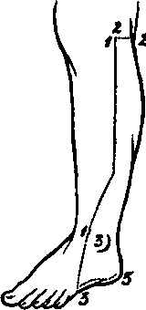
I. Skin Incisions
1. From the head of the Fibula along the upper two thirds of the bone, to the centre of the anterior surface of the ankle-joint.
2. A transverse incision from the upper end of No. 1 backwards, to a little beyond the middle of the posterior surface of the leg.
3. A third from the lower end of No. 1 downwards and forwards to the base of the fifth Metatarsal bone, then backwards along the inner border of the foot to the centre of the heel. Reflect the flap backwards, and expose the superficial fascia and the following parts
No. 22.
1. The triangular subcutaneous surface and the external Malleolus of the Fibula.
2. The External Saphenous vein, from its commencement at the outer side of the Dorsum of the foot, upwards behind the outer ankle and along the median line of the leg through the whole extent of the dissection.
3. Cutaneous arteries.
a. From the Popliteal above and externally, with branches of the External Popliteal nerve.
b. From the Sciatic along the median line, with the Small Sciatic nerve, reinforced by twigs from the Popliteal artery.
c. Numerous branches from the Peroneal along the outer border of the Fibula, and others behind the external Malleolus.
d. From the Anterior Tibial artery in front of the Fibula, with branches of the Musculocutaneous nerve; and the External Malleolar arterial branch in front of the ankle.
e. Twigs from the External Plantar and Tarsal arteries at the outer side of the Dorsum of the foot.
4. Cutaneous branches of the External Popliteal nerve at the upper and outer part; one larger than the rest, called ramus Communicans Fibularis, running downwards and inwards to the middle of the calf.
5. The small portion of the Communicans Tibialis nerve seen superficial to the deep fascia, a little above the centre of the calf.
6. The nerve called External Saphenous resulting from the junction of the preceding, and accompanying the vein of the same name behind the external Malleolus to the outer border of the foot.
7. The Small Sciatic nerve perforating the fascia lata just below the Popliteal space, running down the middle of the leg, and joining the Communicantes Tibialis and Fibularis nerves just at their union. Branches of this nerve are cutaneous at the upper part.
8. In front of the Fibula branches of the Musculocutaneous nerve, which run to the fourth and fifth toes, and other filaments supplying the skin on the outer side of the Dorsum of the foot.
II
Remove the preceding structures and expose the fascia lata, which will be seen forming the upper and lower divisions of the Anterior Annular ligament in front of the ankle, and the External Annular ligament between the Malleolus and Os Calcis. In front of the exposed portion of the Fibula the fascia is very strong, and joined to the outer border of the bone.
III
Remove the above fascial structures and expose
1. The Gastrocnemius muscle and tendo Achillis.
2. The outer border of the Soleus muscle, seen beneath the preceding.
3. The Peroneus longus muscle lying on the Fibula, and covering in the Peroneus brevis muscle except for the anterior part of the lower third.
4. The lower part of the Peroneus tertius muscle in front of the Fibula.
5. The upper part of the origin of the Extensor brevis digitorum muscle on the Dorsum of the foot.
6. The Anterior Peroneal artery and vein in front of the lower end of the Fibula, and on the outer side of the Dorsum of the foot.
7. The small portion of the lesser Sciatic nerve, which, in this dissection, lies beneath the fascia lata.
8. The greater part of the Communicans Tibialis nerve, which is beneath the deep fascia.
9. The External Popliteal nerve at the upper part of the head of the Fibula.
IV
Divide the Oastrocnemius and Soleus muscles above as far as the centre, and pull them well inwards. In doing this, the nerve to the latter will be probably cut through.
Remove the aponeurotic arch over the upper part of the Posterior Tibial vessels and nerve.
There will now be exposed
1. The Popliteus muscle covered by its fascia at the upper part, with its nerve lying on the posterior surface, and turning round the lower border.
2. The aponeurosis stretching over the Tibialis posticus muscle, between the bones of the leg, immediately below it. The fibres of the muscle are visible through the aponeurosis.
3. The Plexor longus pollicis muscle, lying on the lower two thirds of the inner surface of the Fibula.
4. The Popliteal artery and its bifurcation. The Anterior Tibial branch passing between the lower border of the Popliteus muscle and the Tibialis posticus muscle. The Posterior Tibial branch lying on the aponeurosis of the Tibialis posticus muscle, and giving off the Peroneal artery, which runs downwards and outwards for a short distance to enter the substance of the Flexor longus pollicis muscle.
5. The vena comites of the preceding, uniting to form the Popliteal vein.
6. The Posterior Tibial nerve at first to the inner side of the artery of the same name, but soon crossing it to the outer side. The branches to the Tibialis posticus and Flexor longus pollicis muscles are seen, the latter running with the Peroneal artery.
V
a. Trace down the Peroneal artery in the substance of the Flexor longus pollicis muscle, divide the muscle below and pull its lower end downwards. The nutrient and muscular branches will be now seen.
b. Divide the External Annular ligament if not done previously, and open up the sheath of the Pero-neus longus muscle at the outer border of the foot.
e. Cut through the Peronei longus and brevis muscles, and turn the ends upwards and downwards.
d. Saw through the Fibula at the junction of the lower with the upper three fourths, and again two inches lower down, above the external Malleolus. Remove this piece of bone by dividing the attached fibres of the Tibialis posticus and Peroneus tertius muscles and the Interosseous and Inferior Interosseous ligaments. The Peroneal artery may now be followed in its whole course. Below, it is seen lying on the posterior Tibio-fibular ligament, and receiving the communicating branch from the Posterior Tibial artery, which may come over or under the tendon of the Flexor longus pollicis muscle. The artery may be traced down behind the external Malleolus to its anastomoses, with twigs of the Tarsal and External Plantar arteries. The Anterior Peroneal branch may be followed round the front of the outer ankle to its anastomoses with the External Malleolar and Tarsal arteries.
Continue to:
- prev: A Dissection To Expose The Popliteus Muscle
- Table of Contents
- next: A Dissection To Expose The Plantar Arch