The Great Trochanter
Description
This section is from the book "The Anatomy Of The Human Skeleton", by J. Ernest Frazer. Also available from Amazon: The anatomy of the human skeleton.
The Great Trochanter
The great trochanter is a traction epiphysis formed at the attachment of the gluteal muscles, and pulled as a sort of hood over the base of the neck. The descriptive term is applied to the whole mass of bone that stands up at the base of the neck, and does not quite correspond with the epiphysis. The epiphysial line is as shown in Fig. n8, and the epiphysis includes the femoral tubercle at the top of the anterior intertrochanteric line and a part of what is descriptively the upper aspect of the neck.
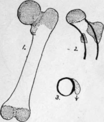
Fig. 116.-1, Diagram of femur at birth ; cartilage is dotted. Upper end shows cartilage to a lower level behind, because the small trochanter is continuous here with great. Observe the short neck. 2, A later condition, in which the shaft (thick lines) has grown in length, carrying the head away from the trochanters and so lengthening the neck. The lower part of the head is formed at first by this shaft, but this is afterwards hidden by epiphysial growth. Trochanters remain as masses applied to the wall of the shaft. 3, A scheme of a section through the small trochanter, showing it applied to the shaft ; the part of the wall which carries it is thick and forms the calcar femorale (see Fig. 125).
The front surface has (Fig. 118) a smooth area for a bursa under the tendon of Gluteus minimus, which is inserted into the strong ridge along its outer border. The, Ilio-trochanteric aponeurosis (p. 124) reaches the ridge below and internal to the bursa. Pyriformis is inserted along the whole length of the upper crest of the trochanter. The strong, direct tendon of the muscle runs mainly into the back half of the crest, where there is usually a facetted appearance made by it and extending back to the unciform end that overhangs the digital fossa.
But this is not the whole attachment of the muscle. If the structures are looked at in an ordinary dissection the tendon of Pyriformis is seen (Fig. 117) to lie in plane superficial to that of the Obturator internus and Gemelli, but if the structures are divided and turned up the Obturator tendon is still seen to be covered by the Pyriformis ; in other words, the Pyriformis partly surrounds the Obturator insertion by means of an expansion passing in front of it, and from this expansion, as shown in the drawing, another aponeurotic sheet is continued to the top of the Gluteus minimus tendon. The complete area of Pyriformis attachment to the bone therefore is as seen in the figure, and the sheet that passes in front of the Obturator tendon is also continued into the fibres of the upper retinaculum.
Obturator internus and Gemelli are inserted internal to and in front of the main tendon of Pyriformis, on the inner side of the trochanter : Gemellus inferior takes the lower and back part of this insertion, and Gemellus superior is largely attached to Pyriformis. The outer surface has a well-marked oblique ridge for the tendon of Gluteus medius, which takes the whole breadth of the ridge. Above and behind this marking may run into that for Pyriformis, when the muscles are partly fused, and in front it is continuous with the lower end of the minimus insertion : these two Glutei are usually fused along their front borders, and it is evident that the fused parts, tying in front of the level of the joint, have similar action as internal rotators.
Above the oblique ridge the trochanter is covered by medius with an intervening bursa, and is thus moulded to a direction differing from that of the bone below the ridge, which is covered only by maximus : this lower surface may have a bursa lying on it, but as a rule the bursa only reaches its most prominent part. The posterior surface is also directly covered by maximus, so that it forms a continuous curve with the area below the oblique ridge.
Obturator externus is not concerned in forming the overhanging trochanter, so that the epiphysial line, which goes to the bottom of the digital fossa (Fig. 118), is further out here than the insertion of the tendon on the floor. The posterior intertrochanteric line is part of the trochanteric system and marks the pull of the Quadratus : this is inserted by muscular fibre as a rule, so that its area is not linear and has not any secondary markings : occasionally there are tendinous fibres, with corresponding secondary roughnesses. The quadrate tubercle is that part of the trochanteric mass on which the pull of the muscle is directly exerted, and it is thus of the nature of a primary marking.
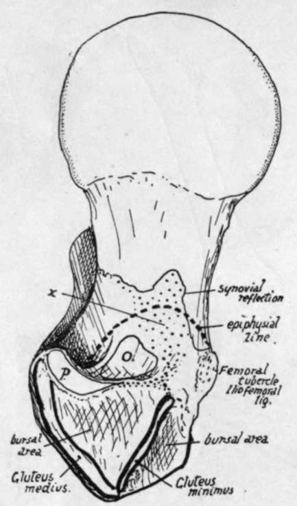
Fig. 117.-Upper aspect of head, neck and great trochanter of right femur. 0. Obturator internus insertion ; P. Pyriformis insertion. The area of insertion of the fibres of the superior retinaculum is marked at x, and it is seen to be continuous with that of Pyriformis round the front of O. and to extend on the neck further than the epiphysial line. Compare with Fig. 118.
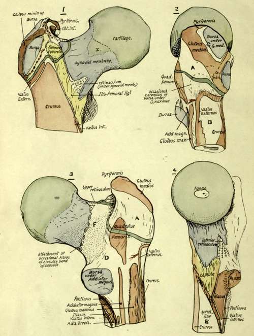
Fig. 118.-Upper end of right femur. The epiphysial line for the great trochanter is marked in green round its base. The retinacula of Weitbrecht," fibres running back toward the head under the synovial membrane, are shown only where they are congregated into their three main groups ; they are derived from the transverse capsular fibres, and the upper one obtains many fibres from Pyriformis (see Fig. 117). I. Anterior aspect. Observe that the Gluteus minimus is attached only to the outer ridge of the trochanter, but its tendon is continuous below with an aponeurotic sheet, the ilio-trochan-teric band, which covers the bursa in front and reaches the bone internal to it. The upper part of the origin of Crureus is mainly tendinous. The extension of the cartilage of the head on to the neck is shown at x ; this lies under the ilio-femoral band or, if the opening for the sub-Psoas bursa is large, under the tendon of the Psoas. 2. From the outer side. The oblique insertion of Gluteus medius is continuous below and in front with that of Gluteus minimus, and frequently with that of Pyriformis above and behind ; it divides this aspect of the trochanter into two areas, one, C, in front and above, under cover of medius and therefore bevelled off in the direction of that muscle, the other, A, below and behind, covered by Gluteus maximus and therefore moulded by that muscle so that it is more vertically directed and curved from before backwards. The surface C carries a bursa, but A has only occasionally an extension of the bursa situated below in relation with it. D, surface covered by Vastus extemus and more or less flattened by it. Crureus fuses with V. externus at a lower level. 3. Posterior aspect. D, surface covered by Quadratus femoris ; deep to this muscle the Obturator externus lies against the bone, moulding the back and lower part of the neck in the area F as it passes to the digital fossa. 4. From the inner side. Observe the pointed area between the spiral line and pectineal line which is occupied by Iliacus. E, inner surface, covered by Vastus internus but not affording origin to it ; the Crureus does not transgress the inner border.
The Small Trochanter is a traction epiphysis at the insertion of the tendon of the Psoas (Fig. 118). The tendon is inserted into its inner rough aspect and, with the Iliacus, into the front surface as well: the Iliacus is also inserted by muscular fibre into the bone in front of and below its level, a triangular area bounded by the spiral and pectineal lines. The back of the small trochanter gives attachment to no muscle, but it is covered by a well-formed bursa which separates it from the top fibres of the Adductor magnus,* into the substance of which it projects when the limb is inverted. This projection can be felt in thin people when the limb is strongly inverted. The plane of the shaft wall deep to the trochanter is strengthened, and forms a bony bar or column visible on section (Fig. 116) and extending up into the neck : this is termed the calcar jemoralc, and is supposed to receive the weight transmitted through the head and from thence by the cancellous arches to the shaft (see Fig. 125). Now articulate the femur and the pelvis and see what the action of the Psoas would be in addition to flexing the joint. Its tendon passes downwards over the front of the joint and turns outwards and backwards to reach its insertion-that is, it pulls on the femur outside the position of the axis of rotation, which passes down through the joint nearly vertically. It is therefore an internal rotator as well as flexor, but the rotating action is probably very small.
Continue to: